Monoclonal Antibodies
Anti-ATP6V0C Rabbit Monoclonal Antibody for WB and ELISA - P27449
Item Number : CM0004716
Price varies based on specs and customizations
- Application
- WB, ELISA
- Cross Reactivity
- Human
- Protein Weight
- 15kDa

Shipping:
Contact us to get shipping details Enjoy On-time Dispatch Guarantee.
Why Choose Us
Easy ordering process, quality products, and dedicated support for your business success.
Core Product Specifications and Parameters
| Parameter | Value |
|---|---|
| Product Name | ATP6V0C Rabbit mAb |
| Remarks/Alias | ATP6V0C; ATP6C; ATP6L; ATPL; VATL; VPPC; Vma3; ATPase H+ transporting V0 subunit c |
| Species | Human |
| Gene ID (Human) | 527 |
| Immunogen | Synthetic peptide; A synthetic peptide corresponding to a sequence within amino acids 1-100 of human ATP6V0C. (NP_001685.1) |
| Source | Rabbit |
| Category | Monoclonal Antibodies |
| Application | WB, ELISA |
| Cross Reactivity | Human |
| SWISS | P27449 |
| Protein Weight | 15kDa |
| Shipping | Ice bag |
Biological Background: ATP6V0C Function and Localization
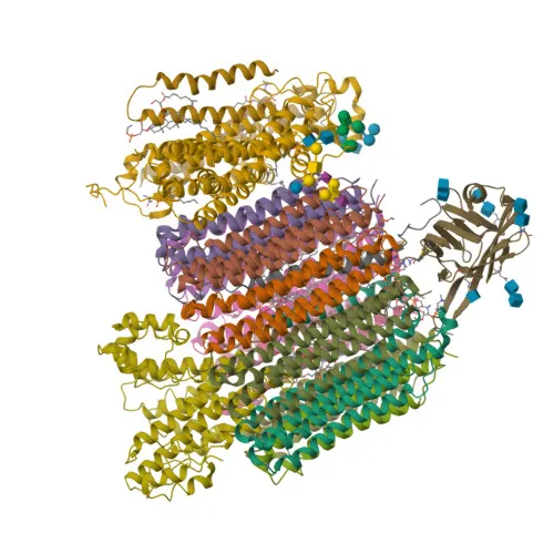
- ATP6V0C encodes the V-type proton ATPase 16 kDa proteolipid subunit c (also known as V-ATPase c subunit), a core component of the V0 membrane-integral complex of vacuolar H+-ATPase (V-ATPase).
- It forms the proton-conducting pore of the V0 complex, facilitating proton translocation across membranes upon ATP hydrolysis by the V1 complex, essential for acidification of intracellular organelles. Related references: PMID:33065002, PMID:36074901
- V-ATPase activity maintains low pH in compartments like lysosomes, endosomes, and secretory vesicles; in some cells, it is also targeted to the plasma membrane to acidify the extracellular milieu.
- The V-ATPase complex, including ATP6V0C, acts as an activator for mTORC1 by promoting the GEF activity of the Ragulator complex on lysosomes, thereby facilitating amino acid-sensing and cell growth signaling. Related references: PMID:22053050
- ATP6V0C is localized to various intracellular membranes: clathrin-coated vesicle membrane, synaptic vesicle membrane, and lysosome membrane, underscoring its role in diverse vesicular acidification processes.
- The protein is subject to ubiquitination, and mutations are associated with epilepsy, highlighting its clinical relevance.
- ATP6V0C is also implicated in host-virus interactions, such as facilitating viral entry or replication in acidic endosomes.
Experimental Guidance and Technical Tips
- The immunogen used is a synthetic peptide corresponding to the N-terminal region (amino acids 1–100) of human ATP6V0C; this region is likely exposed and suitable for detecting the protein in denaturing Western blot conditions.
- For Western blot, the predicted molecular weight is ~15 kDa; ensure adequate separation and transfer for low-molecular-weight proteins.
- For ELISA, consider using the antibody in direct or capture formats depending on the assay design; validate with recombinant ATP6V0C or cell lysates.
- Since cross-reactivity is only confirmed with human, verify species compatibility if using other models.
- As a rabbit monoclonal antibody, it offers high specificity and batch-to-batch consistency; optimize working concentrations empirically for your specific experimental conditions.
CamelBio: Your One-Stop Sourcing Bridge
CamelBio offers comprehensive IVD raw-material sourcing, including validated antibody pairs, optimized monoclonal antibodies like this anti-ATP6V0C rabbit mAb, and bulk ancillary reagents for diagnostic development. Our team supports research on lysosomal acidification, mTORC1 signaling, and vacuolar ATPase function with reliable antibodies and technical consultation. Explore CamelBio’s portfolio to accelerate your assay development from concept to clinic.
REQUEST A QUOTE
Our professional team will reply to you within one business day. Please feel free to contact us!