Polyclonal Antibodies
Anti-ATP6V0A1 Rabbit Polyclonal Antibody for WB, IF/ICC, ELISA - Q93050
Item Number : CM0015621
Price varies based on specs and customizations
- Application
- WB, IF/ICC, ELISA
- Cross Reactivity
- Human, Mouse, Rat
- Protein Weight
- 96kDa

Shipping:
Contact us to get shipping details Enjoy On-time Dispatch Guarantee.
Why Choose Us
Easy ordering process, quality products, and dedicated support for your business success.
Core Product Specifications and Parameters
| Parameter | Value |
|---|---|
| Product Name | ATP6V0A1 Rabbit pAb |
| Remarks/Alias | a1; Stv1; VPP1; Vph1; ATP6N1; DEE104; NEDEBA; ATP6N1A; ATP6V0A1 |
| Species | Human |
| GeneID (human) | 535 |
| GeneID | 535 |
| Immunogen | Recombinant fusion protein containing a sequence corresponding to amino acids 60-300 of human ATP6V0A1 (NP_001123493.1) |
| Source | Rabbit |
| Category | Polyclonal Antibodies |
| Application | WB, IF/ICC, ELISA |
| Cross Reactivity | Human, Mouse, Rat |
| SWISS | Q93050 |
| Protein Weight | 96kDa |
| Shipping | Ice bag |
Biological Background: ATP6V0A1 Function and Localization
-
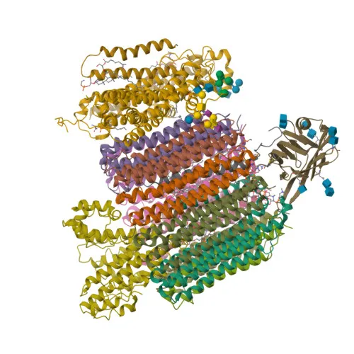
ATP6V0A1 encodes the 116 kDa subunit a1 of the V0 domain of vacuolar H+-ATPase (V-ATPase), a multi-subunit enzyme that pumps protons across cellular membranes. Related references: PMID:33065002, PMID:33833240, PMID:34909687
-
This subunit is essential for proton transport into organelles such as lysosomes, endosomes, the trans-Golgi network, and synaptic vesicles, maintaining acidic luminal pH for protein degradation, receptor trafficking, and neurotransmitter loading.
-
The V-ATPase acts as an activator for mTORC1 on lysosomal membranes by promoting the guanine nucleotide exchange factor (GEF) activity of the Ragulator complex, recruiting mTORC1 to the lysosomal surface. Related references: PMID:22053050
-
In neurons, ATP6V0A1 is critical for neuronal development and connectivity; loss-of-function mutations are associated with developmental and epileptic encephalopathy and intellectual disability. Related references: PMID:33833240
-
The protein localizes to the membrane of clathrin-coated vesicles, synaptic vesicles, and melanosomes, and can also be exported to the plasma membrane in specialized cells to acidify the extracellular environment.
-
ATP6V0A1 undergoes alternative splicing and contains multiple N-glycosylation and phosphorylation sites, which may regulate its stability and function.
Experimental Guidance and Technical Tips
-
Because the immunogen corresponds to a cytoplasmic region (aa 60-300), this polyclonal antibody may effectively detect both native and denatured ATP6V0A1 in Western blot (WB). A band near the predicted 96 kDa may be observed; additional bands from post-translational modifications or isoforms are possible.
-
For immunofluorescence (IF/ICC), the antibody may visualize ATP6V0A1 at intracellular vesicular compartments, such as endosomes and lysosomes. Co-staining with organelle markers can confirm localization.
-
For ELISA, direct or indirect detection of recombinant or purified ATP6V0A1 protein fragments is possible. Titrate the antibody and use appropriate blocking buffers to optimize signal-to-noise.
-
Validate the antibody’s specificity by including positive control lysates (e.g., HeLa, U2OS) and a negative control (knockout or siRNA-treated cells) in your experiments.
CamelBio: Your One-Stop Sourcing Bridge
CamelBio serves as a one-stop sourcing bridge for IVD manufacturers and research laboratories, offering validated antibody pairs, optimized monoclonal/polyclonal antibodies, and bulk ancillary reagents for diverse diagnostic applications. This anti-ATP6V0A1 polyclonal antibody supports research into vacuolar ATPase functions, lysosomal biology, and neurodevelopmental diseases, areas where CamelBio can supply matching reagents and consultative support.
Product Datasheet
Anti-ATP6V0A1 Rabbit Polyclonal Antibody for WB, IF/ICC, ELISA - Q93050
REQUEST A QUOTE
Our professional team will reply to you within one business day. Please feel free to contact us!