Monoclonal Antibodies
Anti-ATP5G1 Monoclonal Antibody for WB, IF-P, ELISA - P05496
Item Number : CM0006674
Price varies based on specs and customizations
- Application
- WB, IF-P, ELISA
- Cross Reactivity
- Human, Mouse, Rat
- Protein Weight
- 14kDa

Shipping:
Contact us to get shipping details Enjoy On-time Dispatch Guarantee.
Why Choose Us
Easy ordering process, quality products, and dedicated support for your business success.
Core Product Specifications and Parameters
| Parameter | Value |
|---|---|
| Product Name | ATP5G1 Rabbit mAb |
| Remarks/Alias | ATP5A; ATP5G; ATP5G1 |
| Species | Human |
| GeneID (Human) | 516 |
| GeneID | 516 |
| Immunogen | A synthetic peptide corresponding to a sequence within amino acids 1-100 of human ATP5G1 (P05496). |
| Source | Rabbit |
| Category | Monoclonal Antibodies |
| Application | WB, IF-P, ELISA |
| Cross Reactivity | Human, Mouse, Rat |
| SWISS | P05496 |
| Protein Weight | 14kDa |
| Shipping | Ice bag |
Biological Background: ATP5G1 Function and Localization
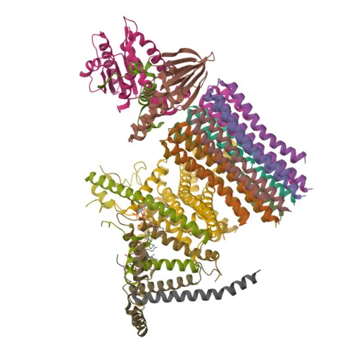
- ATP5G1 encodes ATP synthase F(0) complex subunit C1, also known as ATP synthase lipid-binding protein or ATP synthase membrane subunit c locus 1, a key component of the mitochondrial ATP synthase (Complex V).
- This subunit is an integral part of the F(0) membrane domain, where it collaborates with subunit a (MT-ATP6) to form a proton-conducting channel that translocates protons from the intermembrane space into the mitochondrial matrix, driving ATP synthesis. Related references: PMID:37244256
- The F(0) domain contains two crucial half-channels (inlet and outlet) that facilitate proton movement via a Grotthuss mechanism; proton translocation is coupled to the rotation of the central stalk, linking proton flow to ATP synthesis in the F(1) catalytic head. Related references: PMID:37244256
- ATP5G1 localizes to the mitochondrial inner membrane, as indicated by its transmembrane helices and mitochondrial targeting sequence, ensuring its proper integration into the respiratory chain supercomplex.
- The protein is subject to post-translational methylation, which may regulate its function or assembly within the ATP synthase complex.
- As a core subunit of ATP synthase, ATP5G1 plays a universal role in cellular bioenergetics, making it a valuable marker for mitochondrial function studies across human, mouse, and rat species.
Experimental Guidance and Technical Tips
- The immunogen is a synthetic peptide mapping to the N-terminal region (aa 1-100) of human ATP5G1; for specificity confirmation, consider peptide competition assays using this immunizing peptide alongside full-length recombinant protein.
- In Western blotting, ATP5G1 is expected to appear as a band of ~14 kDa; use mitochondrial-enriched extracts or whole-cell lysates from human, mouse, or rat tissues as positive controls.
- For immunofluorescence (IF-P), thorough cell permeabilization is necessary to detect the mitochondrial membrane antigen; co-staining with mitochondrial markers (e.g., MitoTracker or anti-TOMM20) is recommended to verify localization.
- When performing ELISA, optimize the antibody concentration by titration, and use recombinant ATP5G1 protein or mitochondrial extracts as coating antigen to ensure assay sensitivity.
CamelBio: Your One-Stop Sourcing Bridge
CamelBio serves as a comprehensive sourcing partner for IVD raw materials, offering a wide range of antibodies, antigens, and ancillary reagents for diagnostic development. This anti-ATP5G1 monoclonal antibody is part of our portfolio supporting mitochondrial research and bioenergetics assay development. We provide bulk supply, custom antibody production, and consultative support to accelerate your project from concept to clinic.
REQUEST A QUOTE
Our professional team will reply to you within one business day. Please feel free to contact us!