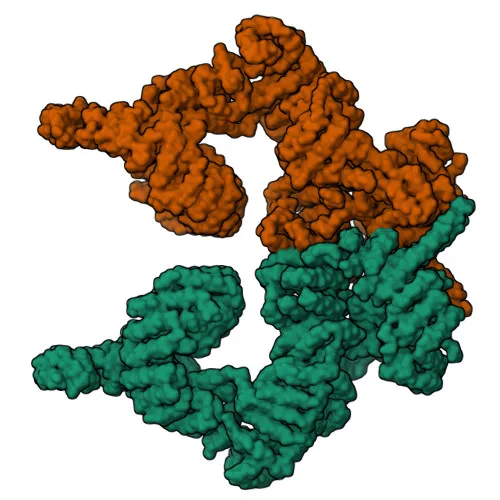
Monoclonal Antibodies
Anti-ATM Monoclonal Antibody for WB, IHC-P, ELISA - Q13315
Item Number : CM0002210
Price varies based on specs and customizations
- Application
- WB, IHC-P, ELISA
- Cross Reactivity
- Human, Mouse
- Protein Weight
- 351kDa

Shipping:
Contact us to get shipping details Enjoy On-time Dispatch Guarantee.
Why Choose Us
Easy ordering process, quality products, and dedicated support for your business success.
Core Product Specifications and Parameters
| Parameter | Value |
|---|---|
| Product Name | ATM Rabbit mAb |
| Remarks/Alias | AT1, ATA, ATC, ATD, ATE, ATDC, TEL1, TELO1, ATM |
| Species | Human |
| GeneID (Human) | 472 |
| GeneID | 472 |
| Immunogen | Recombinant fusion protein containing a sequence corresponding to amino acids 2619-2939 of human ATM (Q13315). |
| Source | Rabbit |
| Category | Monoclonal Antibodies |
| Application | WB, IHC-P, ELISA |
| Cross Reactivity | Human, Mouse |
| SWISS | Q13315 |
| Protein Weight | 351kDa |
| Shipping | Ice bag |
Biological Background: ATM Function and Localization
- ATM (Ataxia telangiectasia mutated) is a serine/threonine protein kinase encoded by the ATM gene, belonging to the PIKK family of kinases involved in DNA damage signaling.
- Functions as a primary sensor of DNA double-strand breaks (DSBs) and genotoxic stress, such as ionizing radiation and ultraviolet A light. It recognizes and phosphorylates substrate proteins on the consensus motif [ST]-Q to initiate checkpoint signaling, apoptosis, and DNA repair. Related references: PMID:10550055, PMID:10839545, PMID:10910365
- Phosphorylates numerous downstream effectors, including histone H2AX (at Ser-139), CHEK2, p53/TP53, BRCA1, and FANCD2, thereby coordinating cell cycle arrest and DNA damage repair pathways. Related references: PMID:10550055, PMID:10766245, PMID:10802669
- Localizes to the nucleus, where it primarily mediates DNA damage signaling, but also resides in cytoplasmic vesicles, centrosomes, and peroxisomes, reflecting its diverse cellular roles.
- Widely expressed in human tissues, with detection in pancreas, kidney, skeletal muscle, liver, lung, placenta, brain, heart, spleen, thymus, testis, ovary, small intestine, colon, and leukocytes.
- Regulates pexophagy by phosphorylating PEX5 at peroxisomes in response to reactive oxygen species, targeting it for ubiquitination and degradation. Related references: PMID:26344566
- Additional functions include enforcing allelic exclusion during B-cell development, controlling replication-dependent histone mRNA degradation, and acting as a potential tumor suppressor.
Experimental Guidance and Technical Tips
- The immunogen corresponds to amino acids 2619–2939 of human ATM, located in the C-terminal region. Validate antibody specificity using appropriate controls, especially when targeting isoforms or splice variants.
- For Western blot, ATM migrates at approximately 351 kDa. Use low-percentage polyacrylamide gels (e.g., 6–8%) and extended transfer times to ensure efficient transfer of this large protein.
- In IHC-P, optimize antigen retrieval methods (e.g., heat-induced epitope retrieval) and primary antibody concentration. Consider using tissues with high ATM expression, such as testis or pancreas, as positive controls.
- Cross-reactivity with mouse is reported; if using mouse models, confirm suitability by testing in relevant knockout or knockdown systems.
- As a monoclonal antibody, it offers high specificity. Include both positive and negative biological controls (e.g., ATM-proficient and ATM-deficient cell lines) to verify band identity.
CamelBio: Your One-Stop Sourcing Bridge
CamelBio offers a comprehensive one-stop sourcing platform for IVD raw materials, from validated monoclonal antibodies like this anti-ATM clone to complementary reagents and custom development services. Focused on DNA damage and cancer research areas, we supply optimized antibody pairs, bulk ancillary reagents, and rare-target raw materials to accelerate diagnostic assay development.
REQUEST A QUOTE
Our professional team will reply to you within one business day. Please feel free to contact us!