Polyclonal Antibodies
Anti-ATL3 Rabbit Polyclonal Antibody for WB, IF/ICC, ELISA - Q6DD88
Item Number : CM0015628
Price varies based on specs and customizations
- Application
- WB, IF/ICC, ELISA
- Cross Reactivity
- Human, Mouse, Rat
- Protein Weight
- 61kDa

Shipping:
Contact us to get shipping details Enjoy On-time Dispatch Guarantee.
Why Choose Us
Easy ordering process, quality products, and dedicated support for your business success.
Core Product Specifications and Parameters
| Parameter | Value |
|---|---|
| Product Name | ATL3 Rabbit pAb |
| Remarks/Alias | HSN1F; ATL3 |
| Species | Human |
| Gene ID (Human) | 25923 |
| Gene ID | 25923 |
| Immunogen | Recombinant fusion protein containing a sequence corresponding to amino acids 360-440 of human ATL3 (NP_056274.3). |
| Source | Rabbit |
| Category | Polyclonal Antibodies |
| Application | WB, IF/ICC, ELISA |
| Cross Reactivity | Human, Mouse, Rat |
| SWISS | Q6DD88 |
| Protein Weight | 61kDa |
| Shipping | Ice bag |
Biological Background: ATL3 Function and Localization
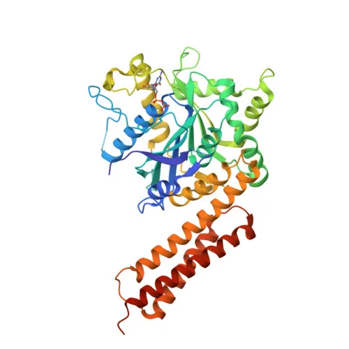
- Atlastin-3 (ATL3), encoded by the ATL3 gene, is a membrane-anchored GTPase belonging to the dynamin-like family; it is also known as HSN1F due to its link with hereditary sensory neuropathy type IF.
- ATL3 mediates GTP-dependent fusion of endoplasmic reticulum (ER) membranes, maintaining the continuous ER network and facilitating the formation of three-way junctions where ER tubules intersect. Related references: PMID:18270207 PMID:19665976 PMID:24459106
- The fusion mechanism involves GTP-induced homodimerization of ATL3 molecules on adjacent ER tubules via their G domains and 3HB regions; GTP hydrolysis triggers conformational changes that pull the membranes together for fusion, after which the dimers disassemble (By similarity).
- Subcellularly, ATL3 is localized to the endoplasmic reticulum membrane, consistent with its role in ER morphogenesis.
- ATL3 is expressed in the central nervous system and dorsal root ganglia neurons, with protein-level detection in peripheral tissues, underscoring its importance in neural ER function.
- Post-translational modifications include acetylation and phosphorylation, and the protein is associated with neuropathy disease variants, ER membrane dynamics, and GTP-binding activity.
Experimental Guidance and Technical Tips
- The immunogen corresponds to amino acids 360–440 of human ATL3, a region likely exposed to the cytosolic side of the ER membrane; consider this when designing epitope retrieval or permeabilization strategies.
- For Western blot, a band around 61 kDa is expected; use appropriate molecular weight markers and validate the antibody in your specific sample system, as expression levels may vary across tissues.
- In IF/ICC, ensure mild permeabilization to maintain ER integrity; co-staining with an ER marker is recommended to confirm localization.
- For ELISA, both direct and indirect formats may be explored; due to the polyclonal nature, lot-to-lot variation may occur, so include a consistent reference standard.
- The antibody shows cross-reactivity with mouse and rat, making it suitable for model organism studies; however, always verify reactivity in your species and tissue of interest.
CamelBio: Your One-Stop Sourcing Bridge
CamelBio serves as a comprehensive sourcing partner for IVD raw materials, technical services, and consulting across the entire diagnostic development pathway. For researchers studying ER dynamics and membrane fusion, we provide access to validated antibody pairs, optimized monoclonal and polyclonal antibodies, bulk ancillary reagents, and rare target raw-material sourcing. Our supply chain expertise supports your ATL3-related projects with consistent quality and reliability.
REQUEST A QUOTE
Our professional team will reply to you within one business day. Please feel free to contact us!