Polyclonal Antibodies
Anti-ATG9B Rabbit Polyclonal Antibody for WB, IF-P, ELISA - Q674R7
Item Number : CM0027013
Price varies based on specs and customizations
- Application
- WB, IF-P, ELISA
- Cross Reactivity
- Human, Mouse, Rat
- Protein Weight
- 101kDa

Shipping:
Contact us to get shipping details Enjoy On-time Dispatch Guarantee.
Why Choose Us
Easy ordering process, quality products, and dedicated support for your business success.
Core Product Specifications and Parameters
| Parameter | Value |
|---|---|
| Product Name | ATG9B Rabbit pAb |
| Remarks/Alias | SONE; APG9L2; NOS3AS; ATG9B |
| Species | Human |
| Gene ID | 285973 |
| Immunogen | Recombinant fusion protein containing a sequence corresponding to amino acids 715-924 of human ATG9B (NP_001303985.1). |
| Source | Rabbit |
| Category | Polyclonal Antibodies |
| Application | WB, IF-P, ELISA |
| Cross Reactivity | Human, Mouse, Rat |
| SWISS | Q674R7 |
| Protein Weight | 101kDa |
| Shipping | Ice bag |
Biological Background: ATG9B Function and Localization
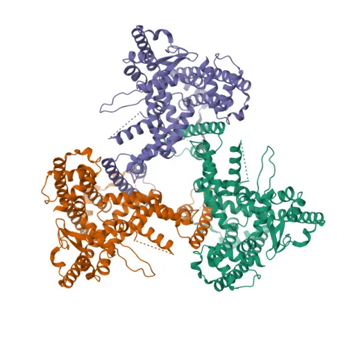
- ATG9B (also known as APG9-like 2 or protein sONE) is a multi-pass transmembrane protein encoded by the ATG9B gene on human chromosome 7.
- It functions as a phospholipid scramblase that plays a critical role in autophagy by mediating autophagosomal membrane expansion.
- The scramblase activity distributes phospholipids from the cytoplasmic to the luminal leaflet of the phagophore, a process that relies on lipid delivery by ATG2 proteins (ATG2A or ATG2B).
- Beyond autophagy, ATG9B is also implicated in necrotic cell death.
- Subcellularly, ATG9B localizes to the membrane of the preautophagosomal structure (phagophore assembly site).
- Tissue expression is predominantly high in placental trophoblast cells and pituitary gland, while absent in vascular endothelial cells.
- The protein belongs to the ATG9 family, and its structure includes multiple transmembrane helices; it is a key component of the autophagy machinery.
Experimental Guidance and Technical Tips
- The immunogen used to generate this antibody corresponds to the C-terminal region (amino acids 715-924) of human ATG9B; the antibody may detect full-length ATG9B as well as any C-terminal fragments.
- For Western blotting, a predicted band around 101 kDa is expected; consider using a 6-8% SDS-PAGE gel for optimal resolution and ensure complete transfer of high-molecular-weight proteins.
- Paraffin-embedded immunofluorescence (IF-P) may require antigen retrieval; validate staining in tissues with known ATG9B expression (e.g., placenta) and include appropriate controls.
- As a polyclonal antibody, lot-to-lot variability is possible; it is recommended to titrate and validate each new lot individually in your specific assay system.
- Cross-reactivity with mouse and rat ATG9B is indicated; confirm sequence homology in the immunogen region for these species if critical.
CamelBio: Your One-Stop Sourcing Bridge
CamelBio provides a one-stop sourcing platform for IVD raw materials, supporting diagnostic developers and researchers with high-quality antibodies, validated antibody pairs, and bulk ancillary reagents. For autophagy and cell death studies, we offer this anti-ATG9B polyclonal antibody alongside custom monoclonal development services to accelerate your assay pipeline. Our supply chain expertise ensures consistent access to rare-target reagents and technical consultation from concept to clinic.
REQUEST A QUOTE
Our professional team will reply to you within one business day. Please feel free to contact us!