Monoclonal Antibodies
Anti-ATG3 Rabbit Monoclonal Antibody for WB, IHC-P, ELISA - Q9NT62
Item Number : CM0002041
Price varies based on specs and customizations
- Application
- WB, IHC-P, ELISA
- Cross Reactivity
- Human, Mouse, Rat
- Protein Weight
- 40kDa

Shipping:
Contact us to get shipping details Enjoy On-time Dispatch Guarantee.
Why Choose Us
Easy ordering process, quality products, and dedicated support for your business success.
Core Product Specifications and Parameters
| Parameter | Specification |
|---|---|
| Product Name | ATG3 Rabbit mAb |
| Remarks/Alias | APG3; APG3L; hApg3; PC3-96; APG3-LIKE |
| Species | Human |
| GeneID (Human) | 64422 |
| Immunogen | Synthetic peptide corresponding to a sequence within amino acids 1-100 of human Apg3 (Atg3) (Q9NT62) |
| Source | Rabbit |
| Category | Monoclonal Antibodies |
| Application | WB, IHC-P, ELISA |
| Cross Reactivity | Human, Mouse, Rat |
| SWISS | Q9NT62 |
| Protein Weight | 40kDa |
| Shipping | Ice bag |
Biological Background: ATG3 Function and Localization
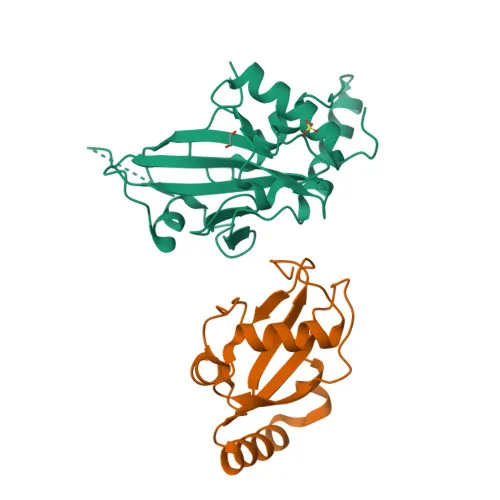
- ATG3 (autophagy-related protein 3, also known as APG3-like, hApg3, and PC3-96) is a ubiquitin-like-conjugating enzyme (E2) essential for autophagy. It catalyzes the covalent conjugation of the C-terminal glycine of ATG8-like proteins (GABARAP, GABARAPL1, GABARAPL2, MAP1LC3A) to phosphatidylethanolamine (PE) on membranes, a key step in autophagosome formation. Related references: PMID:24191030 PMID:33446636 PMID:37252361
- ATG3 cycles between binding to ATG7 for loading with ATG8-like proteins and interacting with the E3 complex (ATG12-ATG5-ATG16L1) to promote lipidation, underpinning the elongation of phagophore membranes. Related references: PMID:11825910 PMID:12207896 PMID:12890687
- It functions as a membrane curvature sensor, detecting lipid-packing defects and enhancing LC3/GABARAP lipidation, and interacts with negatively-charged membranes to promote tethering.
- ATG3 also acts autocatalytically, conjugating ATG12 to itself in an ATG7-dependent manner; this ATG12-ATG3 conjugate regulates mitochondrial homeostasis, late endosome-to-lysosome trafficking, and basal autophagosome maturation.
- Through the autophagic pathway, ATG3 mediates the removal of OFD1 from centriolar satellites, thereby promoting primary ciliogenesis.
- The protein is widely expressed, with highest levels in heart, skeletal muscle, kidney, liver, and placenta, and is subject to acetylation and alternative splicing. Keywords include autophagy, protein transport, and Ubl conjugation pathway.
Experimental Guidance and Technical Tips
- The immunogen is a synthetic peptide from the N-terminus (aa 1–100) of human ATG3, designed to provide high specificity. Researchers should verify recognition of their target isoform or species ortholog.
- For Western blot, a band around 40 kDa is expected; however, post-translational modifications or conjugation events (e.g., ATG12-ATG3) may produce additional bands. Use appropriate controls and validate in your experimental system.
- The antibody is reported to cross-react with mouse and rat ATG3, enabling comparative studies. Confirm cross-reactivity under your specific assay conditions.
- For immunohistochemistry (IHC-P), optimize antigen retrieval and antibody dilution. Negative and positive tissue controls are recommended.
- In ELISA applications, consider pairing with appropriate detection reagents and validate the dynamic range for your analyte.
CamelBio: Your One-Stop Sourcing Bridge
CamelBio provides a one-stop sourcing solution for IVD raw materials, including this ATG3 rabbit monoclonal antibody, serving diagnostic manufacturers, laboratories, and research institutes. With deep expertise in autophagy and cellular clearance pathways, we supply validated antibody pairs, optimized monoclonal/polyclonal antibodies, bulk ancillary reagents, and can source rare-target raw materials to meet your assay development needs.
REQUEST A QUOTE
Our professional team will reply to you within one business day. Please feel free to contact us!