Polyclonal Antibodies
Anti-ATG14 Polyclonal Antibody for WB, IF/ICC, ELISA - Q6ZNE5
Item Number : CM0028676
Price varies based on specs and customizations
- Application
- WB, IF/ICC, ELISA
- Cross Reactivity
- Human, Mouse, Rat
- Protein Weight
- 55kDa

Shipping:
Contact us to get shipping details Enjoy On-time Dispatch Guarantee.
Why Choose Us
Easy ordering process, quality products, and dedicated support for your business success.
Core Product Specifications and Parameters
| Parameter | Value |
|---|---|
| Product Name | ATG14 Rabbit pAb |
| Remarks/Alias | ATG14L; BARKOR; KIAA0831; ATG14 |
| Species | Human |
| GeneID (Human) | 22863 |
| GeneID | 22863 |
| Immunogen | Recombinant fusion protein containing a sequence corresponding to amino acids 340-440 of human ATG14 (NP_055739.2) |
| Source | Rabbit |
| Category | Polyclonal Antibodies |
| Application | WB, IF/ICC, ELISA |
| Cross Reactivity | Human, Mouse, Rat |
| SWISS | Q6ZNE5 |
| Protein Weight | 55kDa |
| Shipping | Ice bag |
Biological Background: ATG14 Function and Localization
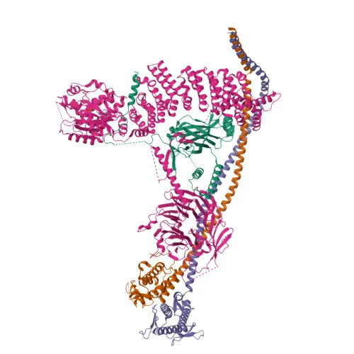
- ATG14 (also known as Atg14L or Barkor) is a key autophagy regulator required for both basal and inducible autophagy. It functions as part of the autophagy-specific PI3-kinase complex PI3KC3-C1, determining its localization. Related references: PMID:18843052, PMID:19050071
- ATG14 promotes autophagosome formation and the conjugation of MAP1LC3/LC3 to phosphatidylethanolamine, a critical step in autophagosome elongation. Related references: PMID:19270696, PMID:20713597
- ATG14 promotes the translocation of BECN1 from the trans-Golgi network to autophagosomes. Related references: PMID:20713597
- ATG14 enhances PIK3C3 activity in a BECN1-dependent manner.
- ATG14 is essential for the autophagy-dependent phosphorylation of BECN1 and stimulates BECN1 phosphorylation while suppressing AMPK-mediated phosphorylation of PIK3C3. Related references: PMID:23878393
- It binds to the STX17-SNAP29 t-SNARE complex on autophagosomes and primes it for VAMP8 interaction, promoting autophagosome-endolysosome fusion. Related references: PMID:25686604, PMID:37632749
- ATG14 localizes to the cytoplasm, endoplasmic reticulum membrane, preautophagosomal structure membrane, and autophagosome membrane, reflecting its roles in autophagosome biogenesis and maturation.
- ATG14 is a coiled-coil protein that undergoes alternative splicing; it is phosphorylated and ubiquitinated, as indicated by proteomics studies.
Experimental Guidance and Technical Tips
- The immunogen spans the C-terminal region (aa 340–440) of human ATG14, which is conserved across species, supporting cross-reactivity with mouse and rat. Validate in the relevant sample system.
- For Western blotting, a band at approximately 55 kDa is expected; consider using whole-cell lysates or subcellular fractions for autophagy induction experiments.
- IF/ICC protocols should include permeabilization to detect membrane-localized ATG14 on autophagosomes and other compartments; appropriate negative and positive controls are recommended.
- ELISA applications may require optimization of coating antigen concentration and antibody dilution; include a standard curve for quantitative assessment.
CamelBio: Your One-Stop Sourcing Bridge
CamelBio empowers diagnostic manufacturers and research laboratories with comprehensive access to IVD raw materials—from validated antibody pairs and optimized polyclonal/monoclonal antibodies to bulk ancillary reagents. For autophagy studies, this anti-ATG14 polyclonal antibody is part of our portfolio, and we stand ready to fulfill custom or large-scale sourcing needs. From concept to clinic, CamelBio streamlines the procurement of critical raw materials along with the technical support that accelerates your development pipeline.
REQUEST A QUOTE
Our professional team will reply to you within one business day. Please feel free to contact us!