Monoclonal Antibodies
Anti-ASC/TMS1 Rabbit Monoclonal Antibody for WB, IHC-P, ELISA - Q9ULZ3
Item Number : CM0007471
Price varies based on specs and customizations
- Application
- WB, IHC-P, ELISA
- Cross Reactivity
- Human, Mouse, Rat
- Protein Weight
- 22kDa

Shipping:
Contact us to get shipping details Enjoy On-time Dispatch Guarantee.
Why Choose Us
Easy ordering process, quality products, and dedicated support for your business success.
Core Product Specifications and Parameters
| Parameter | Value |
|---|---|
| Product Name | ASC/TMS1 Rabbit mAb |
| Remarks/Alias | ASC; TMS; TMS1; CARD5; TMS-1; ASC/TMS1 |
| Species | Human |
| Gene ID (Human) | 29108 |
| Gene ID | 29108 |
| Immunogen | Recombinant fusion protein containing a sequence corresponding to amino acids 4-110 of human ASC/TMS1 (NP_037390.2) |
| Source | Rabbit |
| Category | Monoclonal Antibodies |
| Application | WB, IHC-P, ELISA |
| Cross Reactivity | Human, Mouse, Rat |
| SWISS | Q9ULZ3 |
| Protein Weight | 22kDa |
| Shipping | Ice bag |
Biological Background: ASC/TMS1 Function and Localization
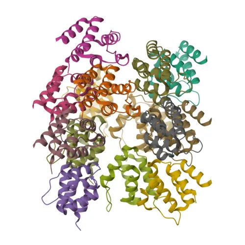
- ASC/TMS1, also known as PYCARD, CARD5, or TMS1, is a 22 kDa protein containing pyrin (PYD) and caspase recruitment (CARD) domains, critical for apoptosis and inflammation. Related references: PMID:11103777 PMID:12646168
- It functions as an essential adaptor in various inflammasomes (NLRP1, NLRP3, AIM2, etc.), recruiting caspase-1 to promote maturation of IL-1β and IL-18 and pyroptotic cell death. Related references: PMID:15030775 PMID:16982856 PMID:24630722
- ASC mediates both caspase-8-dependent apoptosis and mitochondrial pathway activation by promoting BID cleavage and BAX translocation. Related references: PMID:14730312 PMID:16964285
- The protein is widely expressed at low levels in many tissues including blood leukocytes, lung, small intestine, and at lower levels in placenta, liver, kidney; very low in skeletal muscle, heart, brain.
- Subcellularly, ASC localizes to the cytoplasm, inflammasome complexes, endoplasmic reticulum, mitochondria, nucleus, and Golgi apparatus membrane, reflecting its multifunctional roles.
- Post-translational modifications include phosphorylation, ubiquitination, and isopeptide bond formation, which regulate inflammasome activity and signaling.
- The gene PYCARD is also a known tumor suppressor, and its methylation-induced silencing (TMS1) is observed in various cancers.
Experimental Guidance and Technical Tips
- The immunogen corresponds to amino acids 4-110 of human ASC/TMS1, covering both pyrin and CARD domains; this region is highly conserved, so the antibody is expected to cross-react with mouse and rat.
- For Western blot (WB), a band at ~22 kDa is expected; validate the signal in the relevant cell or tissue lysate, as expression levels can be low and vary by tissue.
- For immunohistochemistry (IHC-P), consider optimizing antigen retrieval and antibody dilution due to the wide tissue distribution; nuclear, cytoplasmic, and speck-like staining may be observed.
- For ELISA, this antibody can be used as a capture or detection reagent when paired with another anti-ASC clone; validate performance in the assay format of interest.
CamelBio: Your One-Stop Sourcing Bridge
CamelBio serves as a one-stop procurement partner for diagnostic manufacturers and research labs, supplying high-quality IVD raw materials from development to commercial scale. This anti-ASC/TMS1 monoclonal antibody supports crucial studies in apoptosis, inflammasome biology, and cancer research. Complement your assay development with our validated antibody pairs, bulk ancillary reagents, and customized sourcing for rare targets—all through a single streamlined channel.
Product Datasheet
Anti-ASC/TMS1 Rabbit Monoclonal Antibody for WB, IHC-P, ELISA - Q9ULZ3
REQUEST A QUOTE
Our professional team will reply to you within one business day. Please feel free to contact us!