Polyclonal Antibodies
Anti-ARL2 Polyclonal Antibody for WB, ELISA - P36404
Item Number : CM0029853
Price varies based on specs and customizations
- Application
- WB, ELISA
- Cross Reactivity
- Mouse, Rat
- Protein Weight
- 21kDa

Shipping:
Contact us to get shipping details Enjoy On-time Dispatch Guarantee.
Why Choose Us
Easy ordering process, quality products, and dedicated support for your business success.
Core Product Specifications and Parameters
| Parameter | Value |
|---|---|
| Product Name | ARL2 Rabbit pAb |
| Remarks/Alias | ARFL2; MRCS1; ARL2 |
| Species | Human |
| GeneID (Human) | 402 |
| GeneID | 402 |
| Immunogen | Recombinant protein|Recombinant fusion protein containing a sequence corresponding to amino acids 1-184 of human ARL2 (NP_001658.2). |
| Source | Rabbit |
| Category | Polyclonal Antibodies |
| Application | WB, ELISA |
| Cross Reactivity | Mouse, Rat |
| SWISS | P36404 |
| Protein Weight | 21kDa |
| Shipping | Ice bag |
Biological Background: ARL2 Function and Localization
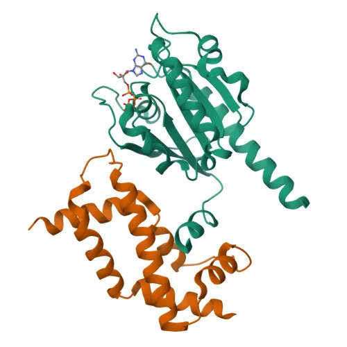
- ARL2 (ADP-ribosylation factor-like protein 2) is a small GTP-binding protein that cycles between inactive GDP-bound and active GTP-bound states, regulated by guanine nucleotide exchange factors (GEFs) and GTPase-activating proteins (GAPs). Unlike ARF proteins, it does not activate cholera toxin catalytic subunit.
- ARL2 is essential for normal cell cycle progression, as it regulates formation of new microtubules and maintains centrosome integrity, also preventing TBCD-induced microtubule destruction.
Related references:
PMID:10831612, PMID:16525022 - In association with TBCD, ARL2 participates in disassembly of apical junction complexes and antagonizes TBCD-mediated epithelial cell detachment and disassembly of tight and adherens junctions.
Related references:
PMID:18234692 - ARL2, together with ARL2, promotes nuclear translocation, retention, and transcriptional activity of STAT3, and is a component of a regulated secretory pathway for Ca(2+)-dependent acetylcholine release.
Related references:
PMID:10831612 - ARL2 regulates mitochondrial integrity and function, as evidenced by recent studies.
Related references:
PMID:30945270 - Subcellularly, ARL2 localizes to the mitochondrial intermembrane space, cytoplasm, cytoskeleton (microtubule organizing center/centrosome), and nucleus.
- Key features include alternative splicing, lipoprotein (myristate), isopeptide bond formation, and phosphorylation; it is linked to cataract when mutated.
Experimental Guidance and Technical Tips
- The immunogen is a recombinant fusion protein covering amino acids 1-184 of human ARL2, nearly the full-length protein; the antibody may detect both GDP- and GTP-bound conformations. Consider testing under conditions that modulate ARL2 activation.
- For Western blotting, a band of approximately 21 kDa is expected. Validate endogenous expression in relevant cell lines or tissues using appropriate positive and negative controls, and confirm specificity with siRNA knockdown or overexpression.
- ELISA can be utilized for detecting human ARL2 in purified recombinant samples or cell lysates; cross-reactivity with mouse and rat makes this antibody suitable for rodent model studies. Optimize coating and detection conditions experimentally.
- Given the mitochondrial and centrosomal localization, immunofluorescence may be attempted after proper fixation and permeabilization; however, this application is not validated and should be empirically tested.
CamelBio: Your One-Stop Sourcing Bridge
CamelBio is a premier supplier of IVD raw materials, offering one-stop access to a comprehensive portfolio that includes high-quality rabbit polyclonal antibodies like anti-ARL2, along with validated antibody pairs, bulk ancillary reagents, and rare target procurement. Supporting research in cell cycle, cytoskeletal dynamics, and mitochondrial biology, we empower diagnostic developers with technical consultation and custom sourcing services to accelerate your concept-to-clinic journey.
REQUEST A QUOTE
Our professional team will reply to you within one business day. Please feel free to contact us!