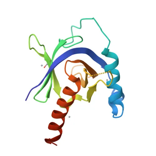
Polyclonal Antibodies
Anti-APBB1 Rabbit Polyclonal Antibody for WB, ELISA - O00213
Item Number : CM0024557
Price varies based on specs and customizations
- Application
- WB, ELISA
- Cross Reactivity
- Human, Mouse
- Protein Weight
- 77kDa

Shipping:
Contact us to get shipping details Enjoy On-time Dispatch Guarantee.
Why Choose Us
Easy ordering process, quality products, and dedicated support for your business success.
Core Product Specifications and Parameters
| Parameter | Value |
|---|---|
| Product Name | APBB1 Rabbit pAb |
| Remarks/Alias | RIR; FE65; MGC:9072; APBB1 |
| Species | Human |
| Gene ID (Human) | 322 |
| Gene ID | 322 |
| Immunogen | Recombinant fusion protein containing a sequence corresponding to amino acids 359-708 of human APBB1 (NP_663722.1) |
| Source | Rabbit |
| Category | Polyclonal Antibodies |
| Application | WB, ELISA |
| Cross Reactivity | Human, Mouse |
| SWISS | O00213 |
| Protein Weight | 77kDa |
| Shipping | Ice bag |
Biological Background: APBB1 Function and Localization
-
APBB1 is a transcription coregulator with both coactivator and corepressor activities, and forms a transcriptionally active complex with the APP intracellular domain. Related references: PMID:15031292, PMID:18468999, PMID:18922798
-
It plays a central role in DNA damage response by recognizing and binding phosphorylated H2AX at Tyr-142 (H2AXY142ph) at double-strand breaks, recruiting pro-apoptotic factors like JNK1, and is required for histone H4 acetylation at DSBs. Related references: PMID:19234442
-
APBB1 specifically binds modified histones and chromatin-modifying enzymes like KAT5/TIP60 to activate transcription, and acts as a transcriptional repressor in complex with TSHZ3, SET, and HDAC to inhibit CASP4 expression. Related references: PMID:33938178, PMID:19343227
-
It localizes to multiple cellular compartments including the cell membrane, cytoplasm, nucleus, growth cones, and nuclear speckles, enabling its diverse signaling and regulatory functions.
-
Highly expressed in the brain, APBB1 levels are reduced in post-mortem Alzheimer disease patients, suggesting a link to neuronal health.
-
It mediates insulin-induced neurite outgrowth by activating the RAC1-ARF6 axis, and also contributes to hippocampal neurite branching, neuromuscular junction formation, lens transparency, and muscle cell strength. Related references: PMID:36250347
-
Post-translational modifications such as phosphorylation, acetylation, and ubiquitin-like conjugation regulate APBB1 activity and interactions.
Experimental Guidance and Technical Tips
- The immunogen corresponds to amino acids 359-708 of human APBB1 (NP_663722.1). For Western blot, consider using a relevant positive control lysate (e.g., human or mouse brain tissue) and validate the observed band against the predicted molecular weight of 77 kDa.
- For ELISA applications, immobilization of the recombinant immunogen or cell lysates may be employed; optimization of coating conditions and antibody dilution is recommended.
- Given cross-reactivity with mouse, this antibody may be suitable for detecting APBB1 in mouse-derived samples; however, species-specific validation in the intended model system is advised.
- As APBB1 is involved in DNA damage response, treatments with genotoxic agents (e.g., etoposide) could be explored to study induced nuclear translocation or post-translational modifications.
CamelBio: Your One-Stop Sourcing Bridge
CamelBio is your one-stop partner for IVD raw-material procurement, offering a comprehensive portfolio that includes validated antibody pairs, monoclonal and polyclonal antibodies, and ancillary reagents. This anti-APBB1 polyclonal antibody, targeting a crucial adapter in Alzheimer's disease and DNA damage signaling, exemplifies our commitment to supplying high-quality reagents for translational research. For bulk orders or custom sourcing of rare-target antibodies, our team provides tailored support to streamline your assay development from concept to clinic.
REQUEST A QUOTE
Our professional team will reply to you within one business day. Please feel free to contact us!