Polyclonal Antibodies
Anti-AP2S1 Rabbit Polyclonal Antibody for WB and ELISA - P53680
Item Number : CM0015431
Price varies based on specs and customizations
- Application
- WB, ELISA
- Cross Reactivity
- Human, Mouse, Rat
- Protein Weight
- 17kDa

Shipping:
Contact us to get shipping details Enjoy On-time Dispatch Guarantee.
Why Choose Us
Easy ordering process, quality products, and dedicated support for your business success.
Core Product Specifications and Parameters
| Parameter | Specification |
|---|---|
| Product Name | AP2S1 Rabbit pAb |
| Remarks/Alias | AP17; FBH3; HHC3; FBHOk; CLAPS2; AP2S1 |
| Species | Human |
| Gene ID (Human) | 1175 |
| Gene ID | 1175 |
| Immunogen | Recombinant protein; Recombinant fusion protein containing a sequence corresponding to amino acids 1-142 of human AP2S1 (NP_004060.2). |
| Source | Rabbit |
| Category | Polyclonal Antibodies |
| Application | WB, ELISA |
| Cross Reactivity | Human, Mouse, Rat |
| SWISS | P53680 |
| Protein Weight | 17kDa |
| Shipping | Ice bag |
Biological Background: AP2S1 Function and Localization
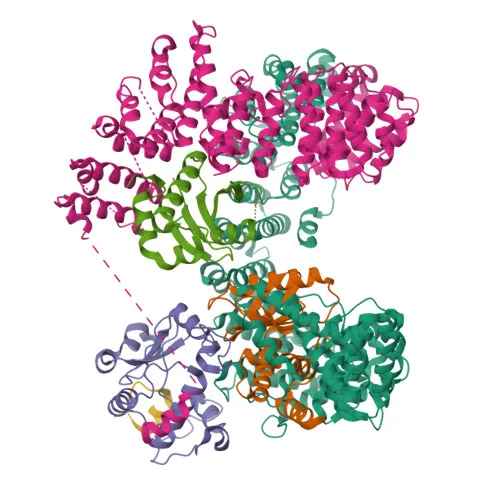
- AP2S1 encodes the sigma subunit of the adaptor protein complex 2 (AP-2), a key component in clathrin-mediated endocytosis.
- AP-2 functions as a vesicle coat component, linking clathrin to membranes and selecting cargo proteins for internalization into clathrin-coated vesicles.
- The complex recognizes specific sorting motifs, such as Y-X-X-Φ and [ED]-X-X-X-L-[LI], within the cytoplasmic tails of transmembrane receptors, enabling selective endocytosis.
- AP-2 sigma and alpha subunits contribute to the recognition of the [ED]-X-X-X-L-[LI] motif.
- AP-2 is also involved in synaptic vesicle membrane recycling from the presynaptic surface.
- Subcellularly, AP2S1 localizes to the cell membrane and coated pits, consistent with its role in vesicle formation.
- The protein is subject to phosphorylation and alternative splicing, and mutations in AP2S1 are associated with disease variants (e.g., in calcium homeostasis disorders).
- Additionally, AP2S1 may play a role in extracellular calcium homeostasis.
Experimental Guidance and Technical Tips
- The immunogen is a recombinant fusion protein corresponding to the N-terminal region (aa 1–142). The antibody may recognize all isoforms of AP2S1.
- For Western blotting, the predicted molecular weight of AP2S1 is ~17 kDa. Validate the antibody using appropriate positive control lysates (e.g., HeLa, HEK293, or tissues rich in endocytic activity).
- In ELISA, the antibody can be used for direct or indirect detection of AP2S1. Titrate the antibody to optimize signal-to-noise ratio.
- Consider testing cross-reactivity in other species if required; provided data show reactivity with human, mouse, and rat.
- Store the antibody as recommended (ice bag shipping implies storage at -20°C or 4°C; avoid repeated freeze-thaw cycles).
CamelBio: Your One-Stop Sourcing Bridge
As a dedicated IVD raw-material supplier, CamelBio offers comprehensive access to antibodies like this AP2S1 polyclonal, which supports research in clathrin-mediated endocytosis and protein transport. Whether you need validated antibody pairs, optimized polyclonal or monoclonal antibodies, bulk ancillary reagents, or assistance in sourcing rare targets, CamelBio streamlines your procurement from concept to clinic. For diagnostic development in endocytosis-related pathways, CamelBio ensures consistent quality and supply chain reliability.
REQUEST A QUOTE
Our professional team will reply to you within one business day. Please feel free to contact us!