Polyclonal Antibodies
Anti-AK2 Polyclonal Antibody for WB and ELISA - P54819
Item Number : CM0026304
Price varies based on specs and customizations
- Application
- WB, ELISA
- Cross Reactivity
- Human, Mouse
- Protein Weight
- 26kDa

Shipping:
Contact us to get shipping details Enjoy On-time Dispatch Guarantee.
Why Choose Us
Easy ordering process, quality products, and dedicated support for your business success.
Core Product Specifications and Parameters
| Parameter | Value |
|---|---|
| Product Name | AK2 Rabbit pAb |
| Remarks/Alias | ADK2; AK2 |
| Species | Human |
| GeneID (Human) | 204 |
| GeneID | 204 |
| Immunogen | Recombinant fusion protein containing a sequence corresponding to amino acids 1-232 of human AK2 (NP_001616.1) |
| Source | Rabbit |
| Category | Polyclonal Antibodies |
| Application | WB, ELISA |
| Cross Reactivity | Human, Mouse |
| SWISS | P54819 |
| Protein Weight | 26kDa |
| Shipping | Ice bag |
Biological Background: AK2 Function and Localization
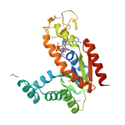
- Adenylate kinase 2 (AK2), also known as ATP-AMP transphosphorylase 2, is a mitochondrial enzyme that catalyzes the reversible transfer of the terminal phosphate group between ATP and AMP, producing two ADP molecules. This reaction is central to cellular energy homeostasis and adenine nucleotide metabolism.
- The adenylate kinase activity is critical for regulating phosphate utilization and the AMP de novo biosynthesis pathways, helping to maintain the appropriate balance of adenine nucleotides within the cell.
- AK2 plays a key role in hematopoiesis, the process of blood cell formation, indicating its importance in hematopoietic stem cell function and differentiation.
- The protein localizes to the mitochondrial intermembrane space, where it participates in the maintenance of the mitochondrial adenine nucleotide pool and energy transfer between mitochondria and the cytosol.
- AK2 is ubiquitously expressed, with high levels in heart, liver, and kidney, and lower levels in brain, skeletal muscle, and skin. Notably, it is present in thrombocytes but absent in erythrocytes, which lack mitochondria. In leukocytes, AK2 is readily detectable, whereas the cytosolic isoform AK1 is mostly absent, suggesting leukocyte susceptibility to AK2 deficiency.
- Post-translational modifications include acetylation and phosphorylation, and the protein contains disulfide bonds and an ATP-binding site. Alternative splicing generates multiple isoforms. Mutations in the AK2 gene are associated with reticular dysgenesis, a severe form of severe combined immunodeficiency (SCID).
Experimental Guidance and Technical Tips
- The immunogen used is a recombinant fusion protein encompassing the full-length sequence (amino acids 1-232) of human AK2. This design may allow the antibody to recognize the native structure of AK2 in various assays. For Western blotting, consider using whole-cell lysates from human or mouse cell lines known to express AK2, such as HeLa or HEK293, and verify the band at ~26 kDa.
- For ELISA applications, the recombinant immunogen can serve as a positive control. Optimization of coating conditions and antibody dilution may be necessary.
- While the antibody has been shown to cross-react with human and mouse AK2, reactivity with other species or isoforms should be validated in the relevant experimental system.
CamelBio: Your One-Stop Sourcing Bridge
At CamelBio, we provide diagnostic manufacturers and research institutions with one-stop access to high-quality IVD raw materials, including validated antibody pairs, recombinant proteins, and ancillary reagents. This AK2 polyclonal antibody supports research into energy metabolism and hematopoiesis, and our team can assist with sourcing rare reagents or scaling up supply for your development needs. From concept to clinic, CamelBio is your trusted partner for reliable raw material procurement and technical support.
REQUEST A QUOTE
Our professional team will reply to you within one business day. Please feel free to contact us!