Polyclonal Antibodies
Anti-AGK Rabbit Polyclonal Antibody for WB, ELISA - Q53H12
Item Number : CM0030337
Price varies based on specs and customizations
- Application
- WB, ELISA
- Cross Reactivity
- Human, Rat
- Protein Weight
- 47kDa

Shipping:
Contact us to get shipping details Enjoy On-time Dispatch Guarantee.
Why Choose Us
Easy ordering process, quality products, and dedicated support for your business success.
Core Product Specifications and Parameters
| Parameter | Value |
|---|---|
| Product Name | AGK Rabbit pAb |
| Remarks/Alias | MULK; CATC5; CTRCT38; MTDPS10; AGK |
| Species | Human |
| Gene ID (Human) | 55750 |
| Gene ID | 55750 |
| Immunogen | Recombinant protein |
| Source | Rabbit |
| Category | Polyclonal Antibodies |
| Application | WB, ELISA |
| Cross Reactivity | Human, Rat |
| SWISS | Q53H12 |
| Protein Weight | 47kDa |
| Shipping | Ice bag |
Biological Background: AGK Function and Localization
- AGK is a mitochondrial lipid kinase that phosphorylates monoacylglycerol and diacylglycerol to produce lysophosphatidic acid (LPA) and phosphatidic acid (PA), respectively. Related references: PMID:15939762
- Overexpression of AGK increases LPA formation and secretion, leading to EGFR transactivation and MAPK pathway activation, thereby promoting cell growth. Related references: PMID:15939762
- AGK also exhibits ceramide kinase activity and preferentially phosphorylates 1,2-dioleoylglycerol over 2,3-dioleoylglycerol (By similarity).
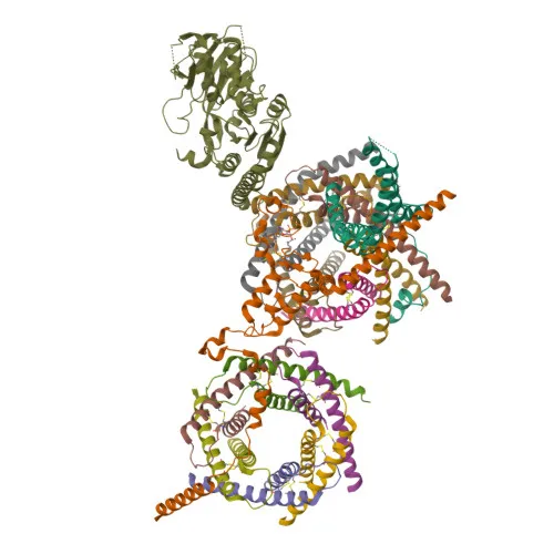
- Independently of its kinase function, AGK is a component of the TIM22 complex in the mitochondrial inner membrane. Related references: PMID:28712724 PMID:28712726
- The TIM22 complex mediates the import and insertion of multi-pass transmembrane proteins, such as metabolite carriers (e.g., ANT1/SLC25A4), into the inner membrane, using the membrane potential as the driving force. Related references: PMID:28712724
- Subcellular localization: mitochondrion inner membrane and intermembrane space.
- Highly expressed in muscle, heart, kidney, and brain.
- Mutations in AGK are associated with diseases including cataract, cardiomyopathy, and primary mitochondrial disorders (Sengers syndrome).
Experimental Guidance and Technical Tips
- The immunogen spans the C-terminal region (aa 233–422) of human AGK, which includes part of the kinase domain. Confirm that the antibody recognizes full-length endogenous AGK in your system.
- For Western blotting, the predicted molecular weight is 47 kDa; validate band size under reducing conditions in relevant tissue or cell lysates.
- AGK is a mitochondrial inner membrane protein, so ensure efficient lysis of mitochondrial membranes (e.g., using RIPA buffer with sonication) for protein extraction.
- The antibody shows cross-reactivity with rat, enabling potential use in rat models; include appropriate positive and negative controls.
- For ELISA, optimize coating antigen concentration and primary antibody dilution using recombinant AGK or cell extracts; consider pairing with a detection antibody from a different host species.
CamelBio: Your One-Stop Sourcing Bridge
CamelBio streamlines IVD raw material sourcing by offering validated antibody pairs, recombinant proteins, and ancillary reagents for mitochondrial and metabolic research. We support diagnostic manufacturers and laboratories with customized polyclonal antibodies, bulk ancillary reagents, and rare target raw materials, enabling efficient development of assays targeting kinases like AGK. Our integrated supply chain and technical consulting help bridge the gap from research concept to clinical application.
REQUEST A QUOTE
Our professional team will reply to you within one business day. Please feel free to contact us!