Monoclonal Antibodies
Anti-AGK Mouse Monoclonal Antibody for WB and ELISA - Q53H12
Item Number : CM0002497
Price varies based on specs and customizations
- Application
- WB, ELISA
- Cross Reactivity
- Human
- Protein Weight
- 47kDa

Shipping:
Contact us to get shipping details Enjoy On-time Dispatch Guarantee.
Why Choose Us
Easy ordering process, quality products, and dedicated support for your business success.
Core Product Specifications and Parameters
| Parameter | Value |
|---|---|
| Product Name | AGK Mouse mAb |
| Remarks/Alias | MULK; CATC5; CTRCT38; MTDPS10; AGK |
| Species | Human |
| GeneID (Human) | 55750 |
| GeneID | 55750 |
| Immunogen | Recombinant fusion protein containing a sequence corresponding to amino acids 1-422 of human AGK (NP_060708.1) |
| Source | Mouse |
| Category | Monoclonal Antibodies |
| Application | WB, ELISA |
| Cross Reactivity | Human |
| SWISS | Q53H12 |
| Protein Weight | 47kDa |
| Shipping | Ice bag |
Biological Background: AGK Function and Localization
- AGK (Acylglycerol kinase, mitochondrial), also known as multiple substrate lipid kinase (HsMuLK), is a dual-function protein.
- As a lipid kinase, it phosphorylates monoacylglycerol and diacylglycerol to produce lysophosphatidic acid (LPA) and phosphatidic acid (PA), respectively. Overexpression enhances LPA secretion, transactivating EGFR and promoting cell proliferation (PubMed:15939762). Related references: PMID:15939762
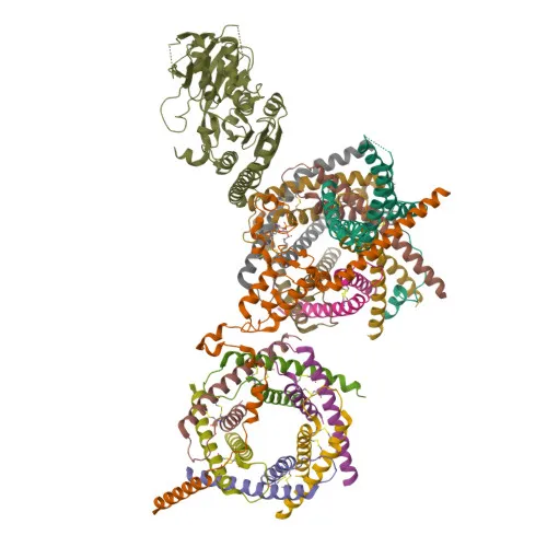
- Independently of its kinase activity, AGK is a crucial subunit of the mitochondrial TIM22 complex, facilitating the import of multi-pass transmembrane proteins like ANT1/SLC25A4 into the inner membrane (PubMed:28712724, PubMed:28712726). Related references: PMID:28712724, PMID:28712726
- Subcellularly, AGK localizes to the mitochondrion inner membrane and intermembrane space.
- AGK is highly expressed in muscle, heart, kidney, and brain.
- Post-translational modifications include acetylation; the protein is also subject to alternative splicing.
- Mutations in AGK are associated with diseases such as Sengers syndrome (cataract and cardiomyopathy) and mitochondrial DNA depletion syndrome.
Experimental Guidance and Technical Tips
- The immunogen covers nearly full-length human AGK (aa 1-422); the antibody likely recognizes both native and denatured conformations, making it suitable for WB under reducing conditions.
- For Western blot, a clear band is expected near 47 kDa. Consider validating in your specific tissue or cell lysate, as expression levels vary (highest in muscle, heart, kidney, brain).
- In ELISA, use the recombinant AGK protein as a standard or capture antigen; pairing with a detector antibody may require optimization of concentrations and buffer conditions.
- Cross-reactivity has been confirmed for human samples only. If working with other species, perform a pilot experiment or check sequence homology, as reactivity cannot be guaranteed without testing.
- As a mouse monoclonal, this antibody is ideal for detection with anti-mouse secondary reagents in various assay formats.
CamelBio: Your One-Stop Sourcing Bridge
CamelBio is a dedicated powerhouse for diagnostic manufacturers, labs, and research institutes, offering streamlined access to premium IVD raw materials, technical services, and expert consulting from concept to clinic. This anti-AGK monoclonal antibody exemplifies our commitment to delivering validated, high-performance research and diagnostic tools for mitochondrial biology and lipid signaling studies. Beyond catalog products, we provide customized solutions including validated antibody pairs, optimized bulk monoclonal/polyclonal antibodies, ancillary reagents, and sourcing for rare targets—all backed by end-to-end support to accelerate your projects.
REQUEST A QUOTE
Our professional team will reply to you within one business day. Please feel free to contact us!