Monoclonal Antibodies
Anti-AGER Rabbit Monoclonal Antibody for WB, IF-P, ELISA - Q15109
Item Number : CM0006575
Price varies based on specs and customizations
- Application
- WB, IF-P, ELISA
- Cross Reactivity
- Human
- Protein Weight
- 43kDa

Shipping:
Contact us to get shipping details Enjoy On-time Dispatch Guarantee.
Why Choose Us
Easy ordering process, quality products, and dedicated support for your business success.
Core Product Specifications and Parameters
| Parameter | Value |
|---|---|
| Product Name | AGER Rabbit mAb |
| Remarks/Alias | RAGE; sRAGE; SCARJ1; AGER |
| Species | Human |
| GeneID | 177 |
| Immunogen | Recombinant protein of human AGER |
| Source | Rabbit |
| Category | Monoclonal Antibodies |
| Application | WB, IF-P, ELISA |
| Cross Reactivity | Human |
| SWISS | Q15109 |
| Protein Weight | 43kDa |
| Shipping | Ice bag |
Biological Background: AGER Function and Localization
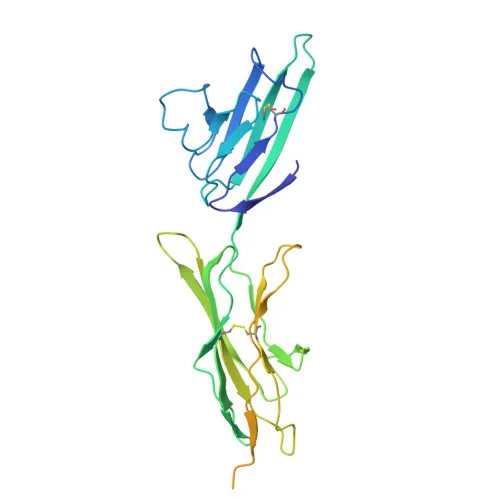
- AGER (also known as RAGE) is a cell surface pattern recognition receptor that binds a broad range of ligands including advanced glycation end products (AGEs), S100 proteins, HMGB1, amyloid beta, nucleic acids, histones, and phospholipids. Related references: PMID:27572515, PMID:28515150, PMID:34743181
- Upon ligand binding, AGER utilizes TIRAP and MYD88 as adaptors to activate NF-kappa-B, leading to the production of pro-inflammatory cytokines such as IL6, IL8, and TNFα. Related references: PMID:21829704, PMID:33436632
- AGER mediates endothelial albumin transcytosis together with HMGB1 via the RAGE/SRC/Caveolin-1 pathway, contributing to endothelial hyperpermeability, and promotes reactive oxygen species (ROS) production in endothelial cells. Related references: PMID:27572515, PMID:25401185
- Following DNA damage, AGER is recruited to double-strand break sites, enhances MRE11 endonuclease activity, promotes DNA damage repair in trophoblasts, and protects cells from replication stress by stabilizing the MCM2-7 complex. Related references: PMID:33918759, PMID:36807739
- AGER promotes non-professional phagocytosis of Saccharomyces cerevisiae spores and apoptotic cells by binding to RNA, DNA, HMGB1, and histones attached to the target. Related references: PMID:35974093
- The receptor is primarily localized at the cell membrane but is also found in phagocytic cups, early endosomes, the nucleus, and can be secreted.
- AGER is highly expressed in endothelial cells, with increased expression noted in pre-term labor and preeclampsia placentas. Related references: PMID:33918759
Experimental Guidance and Technical Tips
- This antibody was raised against recombinant human AGER protein; perform proper controls including recombinant protein competition assays to confirm specificity.
- For Western blot (WB), a band at approximately 43 kDa is expected; use appropriate positive control lysates from endothelial or inflammatory cell lines.
- In immunofluorescence (IF-P), validate staining specificity with knockout or knockdown samples.
- For ELISA applications, titrate the antibody to determine optimal coating and detection concentrations.
CamelBio: Your One-Stop Sourcing Bridge
CamelBio serves as your one-stop IVD raw-material partner, connecting diagnostic manufacturers and research laboratories with high-quality reagents like this anti-AGER antibody for inflammation and diabetes research. We offer validated antibody pairs, optimized monoclonal/polyclonal antibodies, and bulk ancillary reagents to streamline your assay development. Our sourcing expertise extends to even the most niche targets, ensuring you have reliable access to critical raw materials.
REQUEST A QUOTE
Our professional team will reply to you within one business day. Please feel free to contact us!