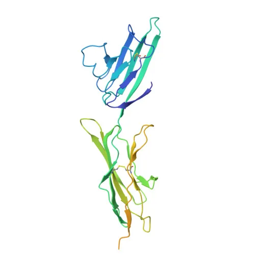
Monoclonal Antibodies
Anti-AGER Monoclonal Antibody for WB - Q15109
Item Number : CM0008675
Price varies based on specs and customizations
- Application
- WB, IF-P, IHC-P, ELISA
- Cross Reactivity
- Human, Mouse, Rat
- Protein Weight
- 43kDa

Shipping:
Contact us to get shipping details Enjoy On-time Dispatch Guarantee.
Why Choose Us
Easy ordering process, quality products, and dedicated support for your business success.
Core Product Specifications and Parameters
| Parameter | Value |
|---|---|
| Product Name | AGER Rabbit PolymAb® |
| Remarks/Alias | RAGE; sRAGE; SCARJ1 |
| Species | Human |
| Gene ID (Human) | 177 |
| Gene ID | 177 |
| Immunogen | Recombinant fusion protein containing a sequence corresponding to amino acids 24-344 of human AGER. (NP_001127.1) |
| Source | Rabbit |
| Category | Monoclonal Antibodies |
| Application | WB, IF-P, IHC-P, ELISA |
| Cross Reactivity | Human, Mouse, Rat |
| SWISS | Q15109 |
| Protein Weight | 43kDa |
| Shipping | Ice bag |
Biological Background: AGER Function and Localization
- AGER (RAGE) is a cell surface pattern recognition receptor that belongs to the immunoglobulin superfamily, playing a central role in innate immunity and inflammation.
- It binds a broad repertoire of endogenous danger signals including advanced glycation end products (AGEs), S100 proteins, HMGB1, amyloid beta, nucleic acids, and histones, triggering pro-inflammatory cascades.
- Upon ligand engagement, RAGE signals via TIRAP and MYD88 adaptors to activate NF-kB, leading to the production of inflammatory cytokines such as IL-6, IL-8, and TNF-α. Related references: PMID:21829704 PMID:33436632
- RAGE also mediates non-professional phagocytosis of apoptotic cells and microbial spores by recognizing surface RNA. Related references: PMID:35974093
- Beyond inflammation, RAGE contributes to DNA damage repair by localizing to double-strand break sites and enhancing MRE11 endonuclease activity, facilitating DNA end resection. (By similarity)
- It promotes DNA replication fork stability by stabilizing the MCM2-7 complex under replication stress, thereby ensuring faithful genome duplication. Related references: PMID:36807739
- Subcellularly, RAGE is found at the cell membrane, phagocytic cup, early endosome, and nucleus, underscoring its multifunctional roles in signal transduction and intracellular trafficking.
- Tissue expression is predominantly in endothelial cells and is elevated in placental pathologies like pre-term labor and preeclampsia. Related references: PMID:33918759
Experimental Guidance and Technical Tips
- The immunogen covers the extracellular domain (aa 24-344) of human AGER, suggesting the antibody may recognize the native receptor and soluble forms (sRAGE).
- For Western blotting, expect a band approximately 43 kDa; validate expression in relevant cell lines or tissues.
- In immunofluorescence and immunohistochemistry, consider permeabilization for intracellular/nuclear pools; antigen retrieval may improve staining in paraffin sections.
- The antibody reacts with mouse and rat AGER, enabling cross-species studies in inflammation and disease models.
- For ELISA applications, test as both capture and detection antibody; pair with appropriate standards for quantitative assays.
CamelBio: Your One-Stop Sourcing Bridge
CamelBio provides diagnostic manufacturers and research laboratories with a comprehensive one-stop shop for IVD raw materials, including validated antibody pairs, optimized monoclonal and polyclonal antibodies, bulk ancillary reagents, and rare target sourcing. This anti-AGER monoclonal antibody supports research into RAGE-mediated inflammation, diabetic complications, neurodegeneration, and related biomarkers. Contact us to discuss your procurement needs, custom formulations, or technical consultation — from concept to clinic.
REQUEST A QUOTE
Our professional team will reply to you within one business day. Please feel free to contact us!