Polyclonal Antibodies
Anti-ADORA1 Polyclonal Antibody for WB, IHC-P, ELISA - P30542
Item Number : CM0020142
Price varies based on specs and customizations
- Application
- WB, IHC-P, ELISA
- Cross Reactivity
- Human, Mouse, Rat
- Protein Weight
- 37kDa

Shipping:
Contact us to get shipping details Enjoy On-time Dispatch Guarantee.
Why Choose Us
Easy ordering process, quality products, and dedicated support for your business success.
Core Product Specifications and Parameters
| Parameter | Value |
|---|---|
| Product Name | ADORA1 Rabbit pAb |
| Remarks/Alias | RDC7; ADORA1 |
| Species | Human |
| Gene ID (Human) | 134 |
| Gene ID | 134 |
| Immunogen | Synthetic peptide|A synthetic peptide corresponding to a sequence within amino acids 250-326 of human ADORA1 (NP_000665.1). |
| Source | Rabbit |
| Category | Polyclonal Antibodies |
| Application | WB, IHC-P, ELISA |
| Cross Reactivity | Human, Mouse, Rat |
| SWISS | P30542 |
| Protein Weight | 37kDa |
| Shipping | Ice bag |
Biological Background: Adenosine receptor A1 Function and Localization
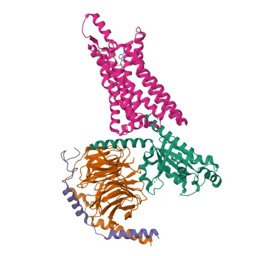
- Adenosine receptor A1 (ADORA1) is a G protein-coupled receptor that binds adenosine. Related references: PMID:29925945
- Activation of ADORA1 triggers inhibitory G proteins such as GNAI2, which inhibit adenylyl cyclase activity, leading to reduced intracellular cAMP levels.
- ADORA1 is localized primarily at the cell membrane and functions as a transmembrane signal transducer.
- The ADORA1 gene undergoes alternative splicing, potentially generating multiple protein isoforms.
- The receptor contains conserved structural motifs typical of the GPCR superfamily, including disulfide bonds, glycosylation sites, and palmitoylation residues.
- ADORA1 has been identified in proteomics studies, indicating its expression in various human tissues.
Experimental Guidance and Technical Tips
- A synthetic peptide from the intracellular C-terminal region (amino acids 250–326) was used as the immunogen; this region is conserved across human, mouse, and rat, supporting the observed cross-reactivity.
- For Western blotting, we recommend using protein lysates from tissues with high ADORA1 expression, such as brain or heart, and optimizing blocking and antibody concentration.
- In immunohistochemistry (paraffin-embedded sections), antigen retrieval conditions should be empirically determined; a positive control tissue with known ADORA1 expression is advised.
- ELISA performance can be influenced by coating antigen and detection system; validate with recombinant or peptide antigen in the relevant buffer system.
- When using this polyclonal antibody, batch-to-batch consistency should be verified for quantitative assays.
CamelBio: Your One-Stop Sourcing Bridge
CamelBio supports diagnostic manufacturers and research laboratories with comprehensive IVD raw material supply, from early R&D to clinical application. This anti-ADORA1 polyclonal antibody addresses GPCR signaling research and assay development needs. Through our global sourcing network, we provide validated antibody pairs, bulk monoclonals and polyclonals, ancillary reagents, and custom procurement for rare targets—delivering reliable, scalable solutions for your workflow.
REQUEST A QUOTE
Our professional team will reply to you within one business day. Please feel free to contact us!