Polyclonal Antibodies
Anti-ACVR2A Rabbit Polyclonal Antibody for WB, IF/ICC, IHC-P, ELISA - P27037
Item Number : CM0022995
Price varies based on specs and customizations
- Application
- WB, IF/ICC, IHC-P, ELISA
- Cross Reactivity
- Human, Mouse, Rat
- Protein Weight
- 58 kDa

Shipping:
Contact us to get shipping details Enjoy On-time Dispatch Guarantee.
Why Choose Us
Easy ordering process, quality products, and dedicated support for your business success.
Core Product Specifications and Parameters
| Parameter | Value |
|---|---|
| Product Name | ACVR2A Rabbit pAb |
| Remarks/Alias | ACVR2; ACTRII; ACVR2A |
| Species | Human |
| GeneID (Human) | 92 |
| GeneID | 92 |
| Immunogen | Recombinant protein |
| Source | Rabbit |
| Category | Polyclonal Antibodies |
| Application | WB, IF/ICC, IHC-P, ELISA |
| Cross Reactivity | Human, Mouse, Rat |
| SWISS | P27037 |
| Protein Weight | 58 kDa |
| Shipping | Ice bag |
Biological Background: ACVR2A Function and Localization
- ACVR2A (Activin receptor type-2A), also known as ACTR-IIA or ACTRIIA, is a transmembrane serine/threonine kinase that belongs to the transforming growth factor-beta (TGF-β) receptor family.
- It functions as a type II receptor for activin A, activin B, and inhibin A. Upon ligand binding, it forms a heteromeric complex with type I receptors, leading to phosphorylation and activation of the type I receptors, which then propagate signals through SMAD transcriptional regulators. Related references: PMID:17911401 PMID:10652306
- ACVR2A mediates the induction of adipogenesis by GDF6, highlighting its role in cellular differentiation processes (by similarity).
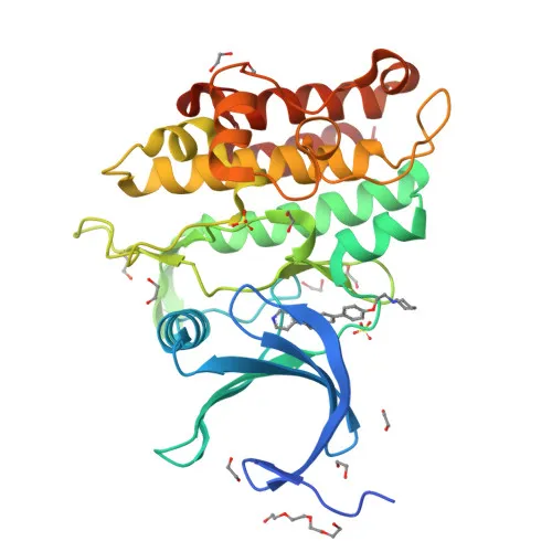
- The protein is localized at the cell membrane, where it acts as a signal transducer, and its extracellular domain contains the ligand-binding region, while the intracellular domain possesses kinase activity.
- Post-translational modifications include glycosylation and disulfide bond formation, which are important for proper folding and function. ACVR2A is also subject to alternative splicing, potentially generating isoforms with distinct regulatory properties.
- Key structural features include an ATP-binding site, a kinase domain, and a single transmembrane helix, with the receptor requiring metal ions such as manganese or magnesium for its enzymatic activity.
Experimental Guidance and Technical Tips
- The immunogen used covers amino acids 20-140 of the extracellular domain, which is expected to generate antibodies recognizing the ligand-binding region; consider this when designing epitope-relevant assays.
- For Western blot (WB), a band around 58 kDa is predicted; however, glycosylation may cause slight migration shifts, so validate in your sample system.
- The antibody is reported to react with human, mouse, and rat species; when using mouse or rat tissues, perform appropriate positive and negative controls to confirm specificity.
- For immunofluorescence/immunocytochemistry (IF/ICC) and immunohistochemistry (IHC-P), cell membrane localization is expected; optimize fixation and permeabilization conditions to preserve epitopes.
- In ELISA applications, pairing with a capture or detection antibody against a non-competing epitope may improve assay performance; test different concentrations to establish optimal conditions.
CamelBio: Your One-Stop Sourcing Bridge
CamelBio provides diagnostic manufacturers, laboratories, and research institutes with one-stop access to IVD raw materials, technical services, and consulting—covering every stage from concept to clinic. For researchers exploring TGF-β/activin signaling and receptor biology, we offer this ACVR2A polyclonal antibody alongside validated antibody pairs, optimized monoclonal/polyclonal antibodies, bulk ancillary reagents, and rare target raw-material sourcing capabilities. Whether your project requires consistent supply or specialized reagents, CamelBio is your trusted B2B partner for IVD raw materials.
Product Datasheet
Anti-ACVR2A Rabbit Polyclonal Antibody for WB, IF/ICC, IHC-P, ELISA - P27037
REQUEST A QUOTE
Our professional team will reply to you within one business day. Please feel free to contact us!