Monoclonal Antibodies
Anti-ACVR2A Rabbit Monoclonal Antibody for WB and ELISA - P27037
Item Number : CM0012045
Price varies based on specs and customizations
- Application
- WB, ELISA
- Cross Reactivity
- Human, Mouse, Rat
- Protein Weight
- 58kDa

Shipping:
Contact us to get shipping details Enjoy On-time Dispatch Guarantee.
Why Choose Us
Easy ordering process, quality products, and dedicated support for your business success.
Core Product Specifications and Parameters
| Parameter | Value |
|---|---|
| Product Name | [KO Validated] ACVR2A Rabbit mAb |
| Remarks/Alias | ACVR2; ACTRII; 2A |
| Species | Human |
| GeneID (human) | 92 |
| GeneID | 92 |
| Immunogen | Synthetic peptide corresponding to a sequence within amino acids 1-100 of human ACVR2A (P27037) |
| Source | Rabbit |
| Category | Monoclonal Antibodies |
| Application | WB, ELISA |
| Cross Reactivity | Human, Mouse, Rat |
| SWISS | P27037 |
| Protein Weight | 58kDa |
| Shipping | Ice bag |
Biological Background: ACVR2A Function and Localization
- ACVR2A encodes a transmembrane serine/threonine kinase receptor for activins and inhibin, key ligands in the TGF-beta superfamily.
- Upon ligand binding, it forms a heteromeric complex with type I receptors, phosphorylating and activating them, which then propagate signals via SMAD transcription factors. Related references: PMID:17911401 PMID:10652306
- It plays a role in adipogenesis, potentially mediated by GDF6 (By similarity).
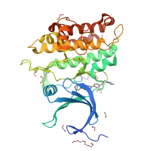
- The receptor contains a signal peptide, extracellular ligand-binding domain, a transmembrane segment, and a cytoplasmic kinase domain.
- Post-translational modifications include glycosylation and disulfide bond formation, essential for proper folding and ligand interaction.
- Alternative splicing yields multiple isoforms, though their functional significance may vary.
- Localizes primarily to the cell membrane, positioning it to interact with circulating ligands.
- Identified as a key node in proteomics studies, underscoring its broad biological relevance.
Experimental Guidance and Technical Tips
- The immunogen corresponds to amino acids 1–100 in the N-terminal extracellular region of ACVR2A (P27037). This may allow the antibody to recognize the receptor’s ligand-binding domain; consider this when designing neutralization studies or receptor detection assays.
- For Western blot (WB), use standard protein extraction from cells or tissues known to express ACVR2A, such as reproductive tissues or skeletal muscle. Validate the band at ~58 kDa.
- Cross-reactivity with mouse and rat has been confirmed; however, individual sample preparation and detection conditions should be optimized for each species.
- In ELISA, include appropriate controls (e.g., blocking peptide or knockout lysates) to confirm specificity, especially when using complex biological matrices.
CamelBio: Your One-Stop Sourcing Bridge
CamelBio provides a streamlined sourcing solution for IVD manufacturers and research labs, offering validated monoclonal antibodies like this ACVR2A rabbit mAb alongside comprehensive raw materials and technical support. From antibody pairs and bulk ancillary reagents to rare target sourcing, we help accelerate your development from concept to clinic. Trust CamelBio as your partner for reliable access to essential tools in TGF-beta signaling and biomarker research.
REQUEST A QUOTE
Our professional team will reply to you within one business day. Please feel free to contact us!