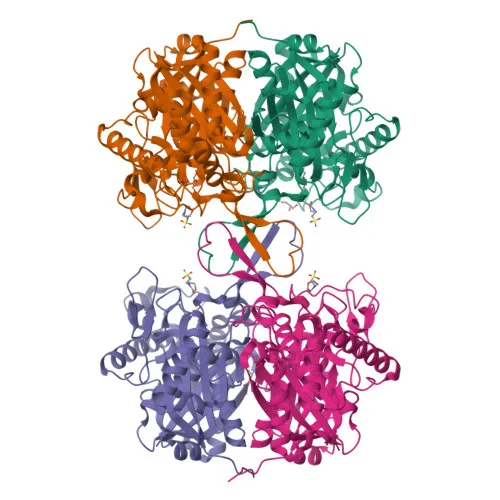
Monoclonal Antibodies
Anti-ACAT1 Rabbit Monoclonal Antibody for WB, IHC-P, IF/ICC, ELISA - P24752
Item Number : CM0007642
Price varies based on specs and customizations
- Application
- WB, IHC-P, IF/ICC, ELISA
- Cross Reactivity
- Human, Mouse, Rat
- Protein Weight
- 45kDa

Shipping:
Contact us to get shipping details Enjoy On-time Dispatch Guarantee.
Why Choose Us
Easy ordering process, quality products, and dedicated support for your business success.
Core Product Specifications and Parameters
| Parameter | Value |
|---|---|
| Product Name | ACAT1 Rabbit mAb |
| Remarks/Alias | T2; MAT; ACAT; THIL; ACAT1 |
| Species | Human |
| Gene ID (Human) | 38 |
| Gene ID | 38 |
| Immunogen | Recombinant protein corresponding to a sequence within amino acids 34-427 of human ACAT1 (NM_000019.4) |
| Source | Rabbit |
| Category | Monoclonal Antibodies |
| Application | WB, IHC-P, IF/ICC, ELISA |
| Cross Reactivity | Human, Mouse, Rat |
| SWISS | P24752 |
| Protein Weight | 45kDa |
| Shipping | Ice bag |
Biological Background: ACAT1 Function and Localization
- ACAT1 encodes mitochondrial acetyl-CoA acetyltransferase, also known as acetoacetyl-CoA thiolase (T2), a crucial enzyme in fatty acid beta-oxidation.
Related references: PMID:1715688, PMID:7728148, PMID:9744475 - It catalyzes the thiolytic cleavage of medium- to long-chain 3-oxoacyl-CoAs into acetyl-CoA and a shortened acyl-CoA, using free coenzyme A, which is the final step of beta-oxidation.
Related references: PMID:1715688, PMID:7728148, PMID:9744475 - The enzyme operates reversibly and can condense two acetyl-CoA molecules to form acetoacetyl-CoA, thus playing a key role in ketone body metabolism.
Related references: PMID:17371050, PMID:1715688, PMID:7728148 - ACAT1 is localized to the mitochondrial matrix.
- Post-translational modifications include acetylation, and the protein undergoes alternative splicing generating multiple isoforms.
- Mutations in ACAT1 are linked to disease variants, emphasizing its importance in metabolic homeostasis.
- Keywords associated with ACAT1 include 3D-structure, metal-binding (potassium), proteomics identification, and reference proteome.
Experimental Guidance and Technical Tips
- For Western blot (WB), a band around 45 kDa is expected. Use appropriate positive controls expressing ACAT1 (e.g., liver or kidney mitochondria extracts).
- For immunohistochemistry (IHC-P), antigen retrieval protocols should be optimized. Mitochondrial staining pattern is anticipated.
- For immunofluorescence (IF/ICC), confirm mitochondrial localization by co-staining with a mitochondrial marker.
- For ELISA, validate linearity and specificity using recombinant ACAT1 protein or target-expressing cell lysates.
- When extending to mouse or rat samples, note that cross-reactivity is reported, but validation in the relevant species is recommended.
CamelBio: Your One-Stop Sourcing Bridge
CamelBio provides diagnostic manufacturers and research laboratories with a comprehensive sourcing platform for IVD raw materials. For metabolic and mitochondrial research, including studies on ACAT1 and fatty acid oxidation, we offer validated monoclonal antibodies, antibody pairs, and bulk ancillary reagents. Our expertise in rare-target sourcing ensures timely access to critical components for assay development.
REQUEST A QUOTE
Our professional team will reply to you within one business day. Please feel free to contact us!