Monoclonal Antibodies
Anti-ACAT1 Rabbit Monoclonal Antibody for WB, IHC-P, ELISA - P24752
Item Number : CM0003587
Price varies based on specs and customizations
- Application
- WB, IHC-P, ELISA
- Cross Reactivity
- Human, Rat
- Protein Weight
- 45kDa

Shipping:
Contact us to get shipping details Enjoy On-time Dispatch Guarantee.
Why Choose Us
Easy ordering process, quality products, and dedicated support for your business success.
Core Product Specifications and Parameters
| Parameter | Value |
|---|---|
| Product Name | ACAT1 Rabbit mAb |
| Remarks/Alias | T2; MAT; ACAT; THIL; ACAT1 |
| Species | Human |
| Gene ID (Human) | 38 |
| Gene ID | 38 |
| Immunogen | Synthetic peptide corresponding to a sequence within amino acids 328-427 of human ACAT1 (P24752) |
| Source | Rabbit |
| Category | Monoclonal Antibodies |
| Application | WB, IHC-P, ELISA |
| Cross Reactivity | Human, Rat |
| SWISS | P24752 |
| Protein Weight | 45kDa |
| Shipping | Ice bag |
Biological Background: ACAT1 Function and Localization
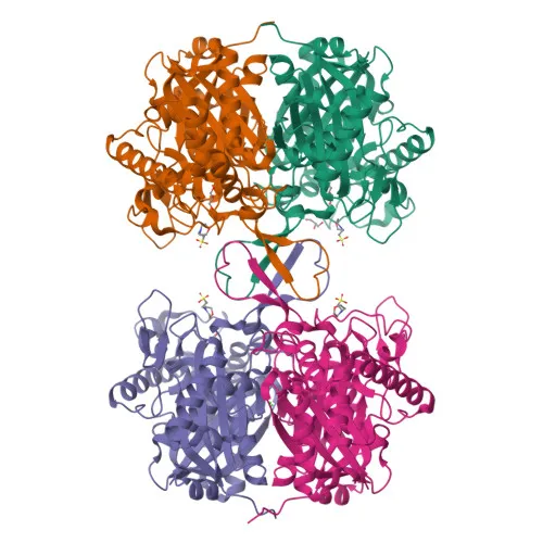
- Acetyl-CoA acetyltransferase, mitochondrial, also known as acetoacetyl-CoA thiolase (T2), is encoded by the ACAT1 gene.
- Catalyzes the last step of mitochondrial fatty acid beta-oxidation: thiolytic cleavage of 3-oxoacyl-CoAs into acetyl-CoA and a shortened acyl-CoA. Reversibly catalyzes the condensation of two acetyl-CoA molecules into acetoacetyl-CoA, playing a major role in ketone body metabolism. Related references: PMID:1715688, PMID:7728148, PMID:9744475, PMID:17371050
- Localized to the mitochondrion, consistent with its central role in fatty acid oxidation and ketogenesis.
- Belongs to the thiolase family of enzymes, which are critical for lipid metabolism.
- Post-translational modifications include acetylation, and the gene undergoes alternative splicing.
- Contains a potassium ion binding site, as indicated by metal-binding properties.
- Mutations in ACAT1 are associated with metabolic disorders, as noted in disease variant annotations.
Experimental Guidance and Technical Tips
- The immunogen corresponds to amino acids 328–427 of human ACAT1, suggesting the antibody recognizes a linear epitope in the C-terminal region.
- In Western blot (WB), a specific band is expected at approximately 45 kDa. Consider using reducing conditions and validate with appropriate positive controls.
- For immunohistochemistry (IHC-P), antigen retrieval may be required; citrate-based methods are commonly used. Optimize antibody dilution and detection conditions.
- ELISA performance may depend on coating antigen and detection format; titration experiments are recommended.
- The antibody cross-reacts with rat ACAT1; validate in the relevant species and tissue systems.
CamelBio: Your One-Stop Sourcing Bridge
CamelBio offers diagnostic manufacturers and research labs seamless access to premium IVD raw materials, including validated antibody pairs and optimized monoclonal/polyclonal antibodies like this anti-ACAT1 clone. Our supply capabilities extend to bulk ancillary reagents and rare target sourcing, supporting projects from discovery to clinical application. For lipid metabolism and metabolic disorder research, we provide integrated procurement solutions to streamline your workflow.
REQUEST A QUOTE
Our professional team will reply to you within one business day. Please feel free to contact us!