Polyclonal Antibodies
Anti-ACAC1/ACC1 Polyclonal Antibody for IHC-P and ELISA - Q13085
Item Number : CM0022721
Price varies based on specs and customizations
- Application
- IHC-P, ELISA
- Cross Reactivity
- Human, Mouse, Rat
- Protein Weight
- 266kDa

Shipping:
Contact us to get shipping details Enjoy On-time Dispatch Guarantee.
Why Choose Us
Easy ordering process, quality products, and dedicated support for your business success.
Core Product Specifications and Parameters
| Parameter | Value |
|---|---|
| Product Name | ACAC1 Rabbit pAb |
| Remarks/Alias | ACC; ACAC; ACC1; ACCA; Acac1; hACC1; ACACAD; ACCalpha; ACACalpha; acetyl-ACC1 |
| Species | Human |
| Gene ID (Human) | 31 |
| Gene ID | 31 |
| Immunogen | Recombinant protein corresponding to a sequence within amino acids 1-75 of human ACAC1 (NP_942133.1) |
| Source | Rabbit |
| Category | Polyclonal Antibodies |
| Application | IHC-P, ELISA |
| Cross Reactivity | Human, Mouse, Rat |
| SWISS | Q13085 |
| Protein Weight | 266kDa |
| Shipping | Ice bag |
Biological Background: ACAC1 Function and Localization
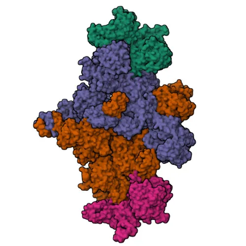
- ACAC1 catalyzes the carboxylation of acetyl-CoA to malonyl-CoA, the first and rate-limiting step of de novo fatty acid biosynthesis. This reaction involves ATP-dependent carboxylation of the biotin prosthetic group followed by carboxyl transfer to acetyl-CoA. Related references: PMID:20457939 PMID:20952656 PMID:29899443
- The enzyme is localized to the cytosol.
- Expressed in brain, placenta, skeletal muscle, renal, pancreatic, and adipose tissues; low in pulmonary tissue; not detected in liver.
- ACAC1 is a metal-dependent enzyme requiring Mg²⁺ or Mn²⁺, and it uses biotin as a covalently attached cofactor.
- Post-translational modifications include phosphorylation and acetylation, which regulate its activity and are identified in proteomics studies.
- As a key enzyme in lipid metabolism, ACAC1 is associated with fatty acid biosynthesis and energy homeostasis.
Experimental Guidance and Technical Tips
- The immunogen targets the N-terminal region (aa 1-75) of human ACAC1. This region may detect full-length ACAC1 or N-terminal truncated variants. Validate specificity in relevant tissue lysates (e.g., adipose or skeletal muscle) using western blot as a complementary technique.
- For IHC-P, optimize antigen retrieval (heat-induced epitope retrieval with citrate buffer, pH 6.0) and primary antibody dilution using known positive samples like adipose tissue or skeletal muscle sections.
- For ELISA, consider using the recombinant immunogen as a positive control and titrate the antibody for optimal signal-to-noise ratio.
- Given cross-reactivity with mouse and rat, the antibody may be suitable for detecting ACAC1 orthologs in these species; verify in respective tissue extracts.
CamelBio: Your One-Stop Sourcing Bridge
As a one-stop IVD raw-material sourcing partner, CamelBio supports diagnostic manufacturers and researchers in the lipid metabolism and metabolic disease fields with high-quality antibodies like this ACAC1 polyclonal antibody. Our catalog includes validated antibody pairs, optimized monoclonal and polyclonal antibodies, and bulk ancillary reagents to accelerate assay development. For rare or custom targets, CamelBio offers flexible sourcing solutions to bridge from concept to clinic.
REQUEST A QUOTE
Our professional team will reply to you within one business day. Please feel free to contact us!