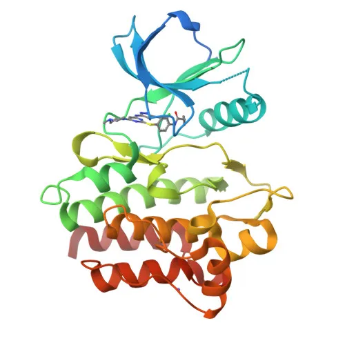
Monoclonal Antibodies
Anti-ABL2 Rabbit Monoclonal Antibody for WB, ELISA - P42684
Item Number : CM0002145
Price varies based on specs and customizations
- Application
- WB, ELISA
- Cross Reactivity
- Human, Rat
- Protein Weight
- 128kDa

Shipping:
Contact us to get shipping details Enjoy On-time Dispatch Guarantee.
Why Choose Us
Easy ordering process, quality products, and dedicated support for your business success.
Core Product Specifications and Parameters
| Parameter | Value |
|---|---|
| Product Name | ABL2 Rabbit mAb |
| Remarks/Alias | ARG; ABLL; ABL2 |
| Species | Human |
| GeneID (Human) | 27 |
| GeneID | 27 |
| Immunogen | A synthetic peptide corresponding to a sequence within amino acids 500-600 of human ABL2 (P42684). |
| Source | Rabbit |
| Category | Monoclonal Antibodies |
| Application | WB, ELISA |
| Cross Reactivity | Human, Rat |
| SWISS | P42684 |
| Protein Weight | 128kDa |
| Shipping | Ice bag |
Biological Background: ABL2 Function and Localization
- ABL2 is a non-receptor tyrosine-protein kinase encoded by the ABL2 gene (also known as ARG, ABLL). It is closely related to ABL1 and contains SH2, SH3, and kinase domains essential for signal transduction.
- It plays a key role in cytoskeleton remodeling by phosphorylating proteins like MYH10, CTTN, TUBA1, and TUBB, and directly binds and bundles F-actin to regulate actin dynamics.
- ABL2 regulates cell adhesion and motility through phosphorylation of CRK, CRKL, DOK1, and ARHGAP35, influencing RHO signaling and the assembly of adhesion complexes.
- It participates in receptor endocytosis by phosphorylating receptor tyrosine kinases such as PDGFRB and endocytic regulators like RIN1.
- In the brain, ABL2 may modulate neurotransmission by phosphorylating synaptic proteins, contributing to neuronal communication.
- It positively regulates chemokine-mediated T-cell migration, polarization, and homing to lymph nodes and immune-challenged tissues, potentially via NEDD9/HEF1 and RAP1.
- Pathogens can subvert ABL2 kinase activity to reorganize the host actin cytoskeleton for intracellular movement and cell exit.
- Subcellularly, ABL2 localizes to the cytoplasm and cytoskeleton. It is widely expressed across tissues, reflecting its broad regulatory roles.
Experimental Guidance and Technical Tips
- The immunogen is a synthetic peptide from human ABL2 (aa 500–600), a region that may be influenced by post-translational modifications or protein interactions; consider epitope accessibility when choosing lysis and detection conditions.
- For Western blotting, expect a band at approximately 128 kDa. Use appropriate reducing and denaturing conditions to resolve the full-length protein, and include molecular weight markers.
- In ELISA, the antibody can be employed as a capture or detection reagent. Optimize coating concentrations and include positive controls (e.g., recombinant ABL2 protein or stimulated cell lysates) for reliable quantification.
- This antibody cross-reacts with rat ABL2, enabling multi-species studies; however, independent validation in the relevant rat tissue or cell line is advised.
- Given ABL2’s involvement in actin dynamics, signal intensity may be enhanced in cells undergoing active cytoskeletal remodeling; consider serum starvation or growth factor stimulation protocols.
CamelBio: Your One-Stop Sourcing Bridge
CamelBio streamlines IVD raw material procurement for diagnostic manufacturers and researchers, offering validated antibody pairs and optimized monoclonal antibodies for areas like cytoskeleton and oncology research. With this ABL2 antibody, along with bulk ancillary reagents and rare target sourcing, CamelBio accelerates development from concept to clinical application.
REQUEST A QUOTE
Our professional team will reply to you within one business day. Please feel free to contact us!