Monoclonal Antibodies
Anti-ABCG1 Rabbit Monoclonal Antibody for WB, ELISA - P45844
Item Number : CM0003002
Price varies based on specs and customizations
- Application
- WB, ELISA
- Cross Reactivity
- Human, Mouse, Rat
- Protein Weight
- 76kDa

Shipping:
Contact us to get shipping details Enjoy On-time Dispatch Guarantee.
Why Choose Us
Easy ordering process, quality products, and dedicated support for your business success.
Core Product Specifications and Parameters
| Parameter | Value |
|---|---|
| Product Name | ABCG1 Rabbit mAb |
| Remarks/Alias | ABC8; WHITE1; ABCG1 |
| Species | Human |
| GeneID (Human) | 9619 |
| GeneID | 9619 |
| Immunogen | Synthetic Peptide |
| Source | Rabbit |
| Category | Monoclonal Antibodies |
| Application | WB, ELISA |
| Cross Reactivity | Human, Mouse, Rat |
| SWISS | P45844 |
| Protein Weight | 76kDa |
| Shipping | Ice bag |
Biological Background: ABCG1 Function and Localization
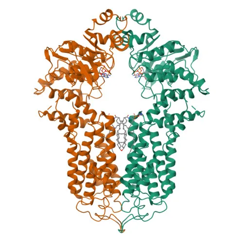
- ABCG1, also referred to as ATP-binding cassette transporter 8 or White protein homolog, is a member of the ATP-binding cassette (ABC) transporter G subfamily and functions as a half-transporter likely forming a homodimer.
- It catalyzes the ATP-dependent efflux of phospholipids such as sphingomyelin, cholesterol, and oxygenated cholesterol derivatives like 7beta-hydroxycholesterol (PubMed:17408620, PubMed:24576892). This lipid transport process is albumin-dependent (PubMed:16702602). Related references: PMID:17408620, PMID:24576892, PMID:16702602
- By exporting cytotoxic 7beta-hydroxycholesterol, ABCG1 prevents cell death, underscoring its role in maintaining cellular lipid homeostasis and protecting macrophages from oxysterol-induced apoptosis (PubMed:17408620). Related references: PMID:17408620
- ABCG1 localizes to the endoplasmic reticulum membrane, Golgi apparatus membrane, and cell membrane, enabling both intracellular lipid trafficking and plasma membrane lipid efflux.
- It is expressed in multiple tissues, with prominent expression in macrophages; notably, ABCG1 expression is upregulated in macrophages from patients with Tangier disease, linking it to cholesterol metabolism disorders.
- Post-translational palmitoylation of ABCG1, as annotated in UniProt, likely modulates its membrane association and functional activity.
- The ABCG1 gene undergoes alternative splicing, generating multiple protein isoforms that may have distinct regulatory or transport properties.
Experimental Guidance and Technical Tips
- The immunogen corresponds to a synthetic peptide derived from amino acids 100–200 of human ABCG1 (NP_004906.3), a region likely exposed in denatured samples, making the antibody potentially suitable for WB under reducing conditions.
- Given the predicted protein weight of 76 kDa, use an appropriate molecular weight marker range and optimize gel percentage (e.g., 8–10% SDS-PAGE) for optimal resolution of ABCG1 bands.
- Cross-reactivity with mouse and rat ABCG1 suggests epitope conservation; validate specificity using lysates from relevant cell types, such as macrophage lines or primary macrophages.
- For ELISA, consider using recombinant ABCG1 protein or the immunizing peptide as a coating antigen and include appropriate positive and negative controls.
- To confirm antibody specificity, incorporate ABCG1-overexpressing cell lysates or knockout/knockdown models and test a range of antibody dilutions.
CamelBio: Your One-Stop Sourcing Bridge
CamelBio serves as a comprehensive sourcing partner for diagnostic manufacturers, laboratories, and research institutes, offering one-stop access to IVD raw materials and technical support from concept to clinic. For studies in lipid metabolism and macrophage biology, we supply a full portfolio of validated antibody pairs, optimized monoclonal/polyclonal antibodies, bulk ancillary reagents, and can assist in navigating rare-target procurement such as ABCG1. Reach out to streamline your supply chain for discovery and diagnostic development.
REQUEST A QUOTE
Our professional team will reply to you within one business day. Please feel free to contact us!